. 1999 Jun;10(6):1821-36.
doi: 10.1091/mbc.10.6.1821.

Amino acid sequence requirements of the transmembrane and cytoplasmic domains of influenza virus hemagglutinin for viable membrane fusion

Affiliations
Free PMC article

Amino acid sequence requirements of the transmembrane and cytoplasmic domains of influenza virus hemagglutinin for viable membrane fusion

G B Melikyan et al. Mol Biol Cell. 1999 Jun.
Free PMC article

Abstract

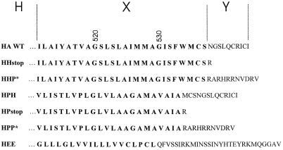
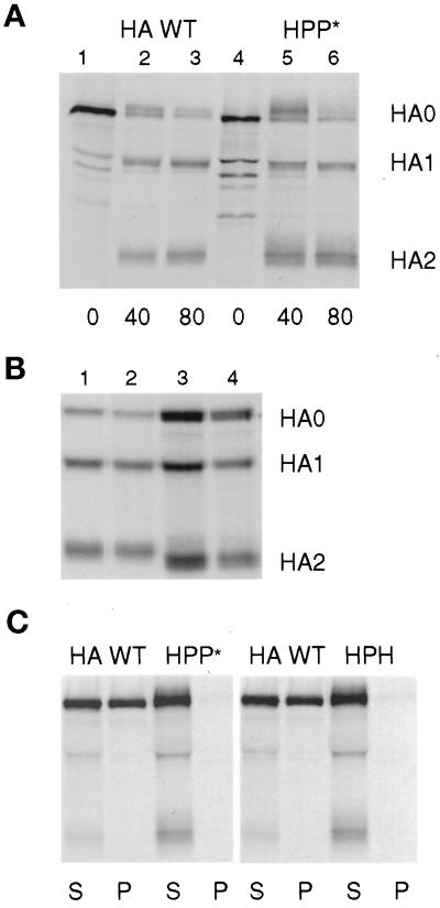
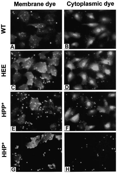
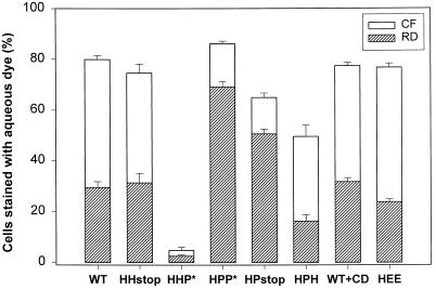
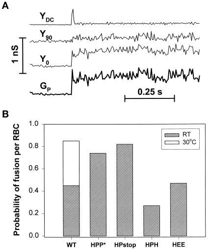
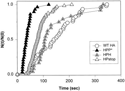
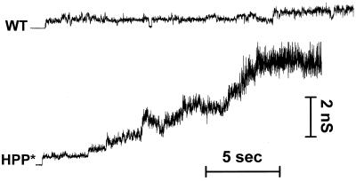
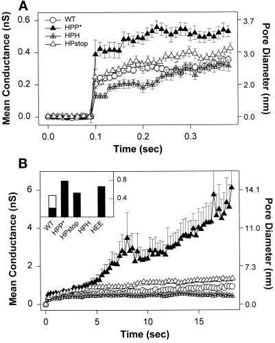
The amino acid sequence requirements of the transmembrane (TM) domain and cytoplasmic tail (CT) of the hemagglutinin (HA) of influenza virus in membrane fusion have been investigated. Fusion properties of wild-type HA were compared with those of chimeras consisting of the ectodomain of HA and the TM domain and/or CT of polyimmunoglobulin receptor, a nonviral integral membrane protein. The presence of a CT was not required for fusion. But when a TM domain and CT were present, fusion activity was greater when they were derived from the same protein than derived from different proteins. In fact, the chimera with a TM domain of HA and truncated CT of polyimmunoglobulin receptor did not support full fusion, indicating that the two regions are not functionally independent. Despite the fact that there is wide latitude in the sequence of the TM domain that supports fusion, a point mutation of a semiconserved residue within the TM domain of HA inhibited fusion. The ability of a foreign TM domain to support fusion contradicts the hypothesis that a pore is composed solely of fusion proteins and supports the theory that the TM domain creates fusion pores after a stage of hemifusion has been achieved.


Figures

Figure 1
Figure 1
Amino acid sequences of the TM domains (bold) and CTs of Japan/305/57 HA WT and HA chimeras constructed for functional studies. The HA constructs are designated H-X-Y, where H is the ectodomain of HA, X is the origin of the TM domain (shown in bold letters), and Y the source of the CT. H denotes HA, P pIgR, and E the env protein of Rous sarcoma virus. A CT of P* symbolizes that the normal CT of pIgR was truncated to 11 amino acid residues to match the length of the CT of HA.
Figure 2
Figure 2
Characterization of the folding and detergent solubility of chimeric HA proteins. (A) CV-1 cells infected with SV40 vectors expressing HA WT or HPP* were pulse labeled for 15 min and chased for the intervals shown in serum-free DMEM containing 10 μg/ml trypsin. Samples were lysed with both Triton X-100 and SDS and immunoprecipitated. HA0, HA1, and HA2 bands were separated by PAGE under reducing conditions. (B) Cells were pulse labeled for 30 min and chased in DMEM for 60 min and then were treated with trypsin in PBS for 60 min at 4°C. Samples were lysed, precipitated, and analyzed as in A. Lane 1, HA WT; lane 2, HHP*; lane 3, HA G530W; lane 4, HA G520L. (C) Cells were labeled with radioactive amino acids for 15 min and chased for 60 min. The cells were then incubated 10 min at 22°C in PBS containing trypsin to cleave HA at the cell surface into HA1 and HA2 subunits. The cells were lysed with Triton X-100 at 4°C, and sample lysates were separated by centrifugation into supernatant (S) and pellet (P) fractions. The pellets were then solubilized by the addition of SDS, and both S and P fractions were immunoprecipitated with antibodies specific for HA. For A–C, digital images of PhosphorImager scans of the dried gels are shown.
Figure 3
Figure 3
Membrane and aqueous dye mixing activity of WT HA and the HEE, HPP*, and HHP* chimeras. All constructs induced R18 to redistribute from RBCs to cells expressing the constructs (left panel). The cytoplasmic marker (CF) transferred efficiently for WT HA and HEE- and HPP*-expressing cells (right panel), demonstrating fusion. In contrast, HHP* only supported limited transfer of a membrane dye without transfer of aqueous dye, suggesting that it caused hemifusion. RBC ghosts colabeled with R18 and CF were bound to HA cells, and fusion was triggered by a 2-min application of pH 4.8 buffer at 37°C, followed by incubation for 5–8 min at neutral pH before microscopically examining cells.
Figure 4
Figure 4
pH dependence of fusion induced by WT and chimeric HA. Fusion between HA-expressing CV-1 cells and RBC ghosts colabeled with CF and R18 was triggered by application of an acidic buffer of the indicated pH for 2 min at 37°C followed by incubation in PBS supplemented with 20 mM raffinose for 5 min at room temperature. For the HHP* construct, the incubation at neutral pH was extended to 15 min. Every point on the graph corresponds to a mean value obtained from four to eight independent experiments (bars indicate SE). The steep pH dependence of WT HA-induced fusion (open circles) was found for all HA chimeras, although the pH curve for the chimeras was shifted by ∼0.2 units toward more acidic pH. Because of the virtual absence of CF transfer for the HHP* construct, its pH dependence is shown for R18 mixing (circles with cross).
Figure 5
Figure 5
Fusion pore enlargement monitored by differential sieving of a large and small cytoplasmic dye. RBC ghosts coloaded with CF (Mr 376) and RD (Mr 40,000) were bound to HA-expressing CV-1 cells, and fusion was triggered by application of a pH 4.8 solution for 2 min at 23°C. Cells were then incubated in PBS with 20 mM raffinose for 5 min (15 min for HHP*) at neutral pH, and the fraction of cells stained by CF (open bars) and RD (striped bars) was determined by fluorescence microscopy. The HHP* chimera promoted virtually no content mixing. Depleting CV-1 cells of cholesterol by incubation with β-cyclodextrin did not affect either the formation or enlargement of WT HA pores. Error bars represent SE for 5–10 independent experiments.
Figure 6
Figure 6
Characteristic electrical signals upon opening of a fusion pore between an RBC and a CV-1 cell and probability that a bound RBC did fuse. (A) The DC conductance (YDC) and in-phase (Y0), and out-of-phase (Y90) conductances were measured by the patch-clamp technique in a whole-cell mode, allowing the fusion pore conductance (GP) to be calculated. The opening of a fusion pore was accompanied by a spike in DC conductance and changes in Y0 and Y90. (B) The number of current spikes in YDC yields the number of RBCs that have fused to the cell. By counting the number of RBCs bound to the CV-1 cell, the probability of fusion per bound RBC was obtained.
Figure 7
Figure 7
Kinetics of fusion between RBCs and HA cells. Fusion was triggered by exposing cells to a pH 4.8 solution for 2 min at room temperature. The waiting times until pore formation were determined electrically. These times were ranked for each construct and plotted as the cumulative distribution of the fraction of cells that have fused by time [i.e. N(t) of the total experimental pool of N(0)]. The kinetics of fusion was similar for WT HA (open circles) and HPH (triangles with cross), each slower than for HPP* (filled triangles) and HPstop (open triangles).
Figure 8
Figure 8
Representative patterns of the evolution of conductance of fusion pores formed by WT HA and HPP*. The conductance much more readily increased for HPP*, and pores did not flicker as much as for WT HA. Experiments were performed at room temperature.
Figure 9
Figure 9
Average conductance profiles of pores at early and later times after formation. Pores were aligned at their opening, and the mean and SE of the conductance over time were determined for the entire population of pores. Sample sizes: WT HA (open circles), n = 32; HPP* (filled triangles), n = 20; HPstop (open triangles), n = 28; and HPH (triangles with cross), n = 27. The approximate diameters of fusion pores in nanometers (Spruce et al., 1989) are shown on the right axis. (A) The initial conductance of pores induced by HPP* was statistically (p < 0.001) larger than formed by the other constructs. (B) Pores formed by HPP* readily enlarge. Once a pore had increased beyond the measurable range, the conductance for this experiment was artificially fixed at the largest detectable value for purposes of calculating the remainder of the profile. This would cause the calculated average conductance to increasingly underestimate the true value as time advances (see MATERIALS AND METHODS). For visual clarity, one of every 25 points of pore profiles is shown. Inset, The fraction of pores that had enlarged beyond 4 nS in 2 min (error bars are omitted for visual clarity). The enlargement for WT HA-expressing cells at 30°C (open bar, n = 14) was greater than at room temperature (filled bars).
Figure 10
Figure 10
Fusion activity of the point mutants G520L HA, G520S HA, and G530W HA. Fusion of bound RBCs colabeled with CF and R18 to HA cells was triggered by application of a pH 4.8 solution at 37°C for 2 min and then incubating the cells for 10 min at room temperature in PBS supplemented with raffinose. The G530W mutant induced efficient redistribution of both dyes (A and B). G520S HA was also effective in promoting fusion (C and D). Very little lipid dye mixing (E) and virtually no aqueous dye redistribution (F) was observed for the G520L mutant. Subsequent application of 0.5 mM CPZ to the G520L-expressing cells resulted in extensive transfer of both CF (H) and R18 (G). The electrical trace (Y0) illustrates that pores did not form for G520L after the low-pH pulse (downward arrow). However, a rare transient opening of a fusion pore followed by its closing was sometimes detected when the probability of pore formation was increased by binding many RBCs to the G520L cells.
Figure 11
Figure 11
Schematic representation of the progression through intermediates in HA-mediated fusion. Each monomer of the HA homotrimer consists of HA1 (shown as a globular head with a short “tail”) and HA2 (a membrane-anchored polypeptide) subunits. The fusion peptides (small open ellipses) are initially sequestered from aqueous phase. Each TM domain is represented by a larger ellipse, and a CT is shown by thin lines. The HA-expressing cell binds to its target membrane (Binding) via specific interactions between the HA1 subunit and sialates on the target membrane. Low pH triggers a series of conformational changes in HA that result in insertion of fusion peptides into viral and target membranes. Fusion probably proceeds from a local membrane merger (Local hemifusion) mediated by the ectodomain of HA. The question mark between the bound and hemifused states denotes that the manner by which HA causes this initial hemifusion is not known. The addition of CPZ converts the local hemifusion intermediate to a pore that has irreversibly opened (Irreversible opening of a fusion pore), effectively bypassing the small flickering pore (Reversible opening of a fusion pore). We propose that for membranes in the state of local hemifusion the combination of the TM domain and CT causes the destabilization of the hemifusion diaphragm that results in an initial pore that can still close. After some enlargement the pore remains open and eventually fully enlarges (Enlarged pore). Stages up to local hemifusion do not require the presence of a TM domain, whereas pore formation does.

Similar articles

Cited by

References

    1. Bagai S, Lamb RA. Truncation of the COOH-terminal region of the paramixovirus SV5 fusion protein leads to hemifusion but not complete fusion. J Cell Biol. 1996;135:73–84. - PMC - PubMed
    1. Blumenthal R, Sarkar DP, Durell S, Howard DE, Morris SJ. Dilation of the influenza hemagglutinin fusion pore revealed by the kinetics of individual cell-cell fusion events. J Cell Biol. 1996;135:63–71. - PMC - PubMed
    1. Brown DA, London E. Structure of detergent-resistant membrane domains: does phase separation occur in biological membranes? Biochem Biophys Res Commun. 1997;240:1–7. - PubMed
    1. Bullough PA, Hughson FM, Skehel JJ, Wiley DC. Structure of influenza hemagglutinin at the pH of membrane fusion. Nature. 1994;371:37–43. - PubMed
    1. Chakrabarti L, Emerman M, Tiollais P, Sonigo P. The cytoplasmic domain of simian immunodeficiency virus transmembrane protein modulates infectivity. J Virol. 1989;63:4395–4403. - PMC - PubMed

Publication types

MeSH terms

Substances

LinkOut - more resources