. 1999 Jun;10(6):1973-84.
doi: 10.1091/mbc.10.6.1973.

The PDZ domain of the LIM protein enigma binds to beta-tropomyosin

Affiliations
Free PMC article

The PDZ domain of the LIM protein enigma binds to beta-tropomyosin

P M Guy et al. Mol Biol Cell. 1999 Jun.
Free PMC article

Abstract

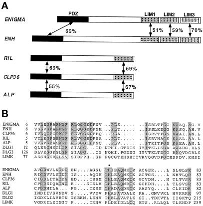
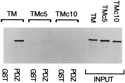
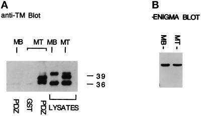
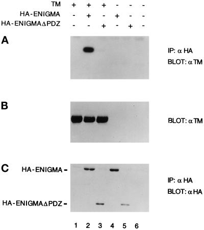
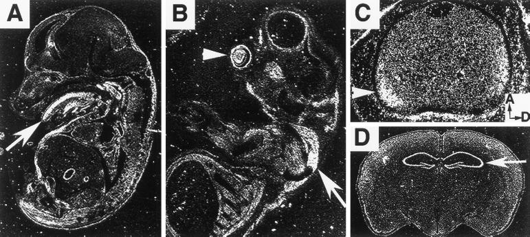
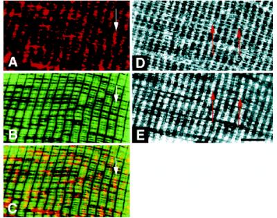
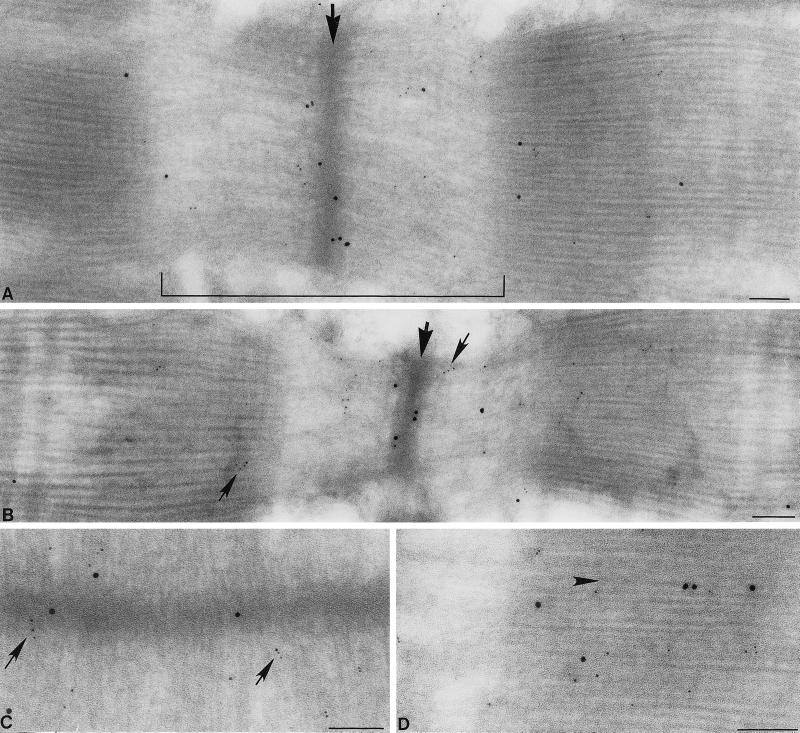
PDZ and LIM domains are modular protein interaction motifs present in proteins with diverse functions. Enigma is representative of a family of proteins composed of a series of conserved PDZ and LIM domains. The LIM domains of Enigma and its most related family member, Enigma homology protein, bind to protein kinases, whereas the PDZ domains of Enigma and family member actin-associated LIM protein bind to actin filaments. Enigma localizes to actin filaments in fibroblasts via its PDZ domain, and actin-associated LIM protein binds to and colocalizes with the actin-binding protein alpha-actinin-2 at Z lines in skeletal muscle. We show that Enigma is present at the Z line in skeletal muscle and that the PDZ domain of Enigma binds to a skeletal muscle target, the actin-binding protein tropomyosin (skeletal beta-TM). The interaction between Enigma and skeletal beta-TM was specific for the PDZ domain of Enigma, was abolished by mutations in the PDZ domain, and required the PDZ-binding consensus sequence (Thr-Ser-Leu) at the extreme carboxyl terminus of skeletal beta-TM. Enigma interacted with isoforms of tropomyosin expressed in C2C12 myotubes and formed an immunoprecipitable complex with skeletal beta-TM in transfected cells. The association of Enigma with skeletal beta-TM suggests a role for Enigma as an adapter protein that directs LIM-binding proteins to actin filaments of muscle cells.


Figures

Figure 1
Figure 1
The Enigma family of PDZ-LIM proteins. (A) Enigma, ENH, RIL, CLP36, and ALP have conserved PDZ and LIM domains and represent a family of PDZ–LIM proteins. Percentages of amino acid identities between LIM and PDZ domains of family members are indicated. (B) Amino acid sequence alignment of PDZ domains of Enigma, ENH, RIL, CLP36, ALP, Dlg (PDZs 1 and 2), and LIM kinase. Four residues that reside in the carboxylate-binding loop of PDZ domains, the Gly-Leu-Gly-Phe sequence for which PDZ domains were originally named, are boxed. Among Enigma family members the sequence (Pro/Ser)-Trp-Gly-Phe occurs at this site. A histidine residue that is conserved in the Enigma family (His-63 in Enigma) is indicated with an asterisk. Amino acid identities among Enigma family and other PDZ domains are indicated by shading. The amino acid sequences used in this study were obtained from the following database entries: Enigma, L35240; ENH, U48247; RIL, X76454; CLP36, U23769; Dlg, M73529; LIM kinase, D26309; and ALP (Xia et al., 1997).
Figure 2
Figure 2
Specificity of the interaction of the PDZ domain of Enigma with TM-1. (A) Detergent extracts of skeletal β-TM fusion protein (35S-T10-TM) generated by in vitro translation were probed with glutathione-agarose–coupled GST or GST-PDZ domains of Enigma, Dlg (PDZs 1 and 2), LIM kinase, and ENH. T10-TM binds selectively to the PDZ domain of Enigma. A Coomassie blue–stained gel of the various GST-PDZ domains is shown in the lower panel. Input represents 2% of the T10-TM extract incubated with GST-PDZ domains. (B) Demonstration of the inability of GST-PDZEng mutants His63Ala and Gly15Ala/Phe16Ala to interact with T10-TM. Input represents 1.5% of the T10-TM extract incubated with GST-PDZ domains. (C) Untagged TM-1 binds to the PDZ domain of Enigma to the same extent as the corresponding fusion protein. Input represents 2% of the TM-1 extract incubated with GST-PDZ domains.
Figure 3
Figure 3
Interaction of the PDZ domain of Enigma requires the carboxyl terminus of skeletal β-TM. Truncation mutants having deletions of 5 or 10 amino acids from the carboxyl terminus of skeletal β-TM (TMc5 and TMc10, respectively) were translated in vitro as T10-TM fusion proteins and incubated with GST-PDZEng or GST alone. Mutant skeletal β-TM proteins did not bind to GST-PDZEng. Input represents 2% of translation reactions of skeletal β-TM, TMc5, and TMc10 incubated with GST beads.
Figure 4
Figure 4
Association of the PDZ domain of Enigma with TM isoforms in C2C12 myotubes. (A) Detergent extracts of C2C12 myoblasts (MB) or myotubes (MT) were incubated with GST or GST-PDZEng immobilized on glutathione-agarose. Two predominant forms of TM were detected in C2C12 lysates by Western blotting with anti-TM antibodies: 40- and 36-kDa forms were present in myoblasts, whereas myotubes expressed 39- and 36.5-kDa form. GST-PDZEng selectively bound to the myotube-specific 39- and 36.5-kDa TM isoforms. Lysates represent 5% of the input cell extract before incubation with GST beads. (B) Anti-Enigma Western blot showing similar levels of Enigma expression in myoblasts (MB) and myotubes (MT).
Figure 5
Figure 5
Coimmunoprecipitation of HA-Enigma and skeletal β-TM in transfected 293 cells. Proteins were expressed via transient transfection of 293 cells using the indicated pcDNA3 constructs: lane 1, skeletal β-TM; lane 2, skeletal β-TM and HA-tagged full-length Enigma; lane 3, skeletal β-TM and HA-tagged Enigma LIM domains (HA-EnigmaΔPDZ); lane 4, HA-tagged full-length Enigma; lane 5, HA-EnigmaΔPDZ; and lane 6, empty vector. Antibodies against the HA epitope were used to coprecipitate HA-Enigma with associated proteins. (A) Anti-TM Western blot indicates that skeletal β-TM associates with HA-Enigma (lane 2) but not with HA-EnigmaΔPDZ (lane 3). (B) Anti-TM Western blot of transfected cell lysates before immunoprecipitation (2% of input). (C) The blot in A was stripped and reprobed with anti-HA antibodies. Bands at ∼56 and ∼25 kDa indicate that both HA-Enigma (lanes 2 and 4) and HA-EnigmaΔPDZ (lanes 3 and 5) were immunoprecipitated by the anti-HA antibodies.
Figure 6
Figure 6
Expression of Enigma in skeletal muscle, brain, and other tissues. (A) In situ hybridization of an E13.5 mouse embryo sagittal section shows Enigma mRNA expression in skeletal muscle. The arrow indicates Enigma expression within the intrinsic and extrinsic muscle mass of the tongue. (B) Expression is observed in epaxial, intercostal, and other skeletal muscles at the brachial level, including the latissimus dorsi muscle, indicated by the large arrow. Lens tissue is indicated by an arrowhead. (C) Higher magnification of lens tissue shown in B. The arrow marks the location of Enigma-expressing lens cells adjacent to the equatorial zone of proliferation. For orientation, anterior (A) and dorsal (D) axes are indicated in the inset. (D) Enigma mRNA is evident within the adult hippocampus, as indicated by the arrow.
Figure 7
Figure 7
Localization of Enigma in skeletal muscle. (A) Immunofluorescence of semithin cryostat sections shows that Enigma is present at the Z line and some transverse filaments in adult muscle sarcomeres. Enigma is present along the Z line as indicated by the arrow. (B) Phalloidin-FITC staining of the same tissue section as in A shows the distribution of actin at the I band and at the Z line, which lies at the midline of the I band. The arrow marks the actin-rich Z line. (C) Digitally merging of the images of A and B shows codistribution of Enigma (red) and Phalloidin-stained actin (green). Regions where Enigma and actin overlap appear yellow, as indicated by the arrow. Arrows in A–C mark the same location in the tissue section. (D) Enigma is detected at the Z line, as indicated by the arrow, and along a subset of transverse filaments. (E) Skeletal β-TM is also distributed near the Z line (arrowhead) in a doublet pattern on the same tissue section as in D. Bar, ∼3 μM.
Figure 8
Figure 8
Immunoelectron microscopic analysis of the distribution of Enigma in skeletal muscle. Immunoelectron micrographs show the presence of Enigma (10-nm gold) and skeletal TM (5-nm gold) in ultrathin sections of adult muscle. (A–C) Enigma (large black dots) is predominantly detected at the boundary of the I band and Z line, whereas skeletal TM (small dots) is detected at the boundary of the I band and Z line and throughout the I band. The arrow in A indicates the Z line, and the bracket spans the I band. The large arrow in B marks the Z line, and small arrows mark skeletal TM detected in the I band and along thin filaments. Arrows in C indicate skeletal TM located at the boundary between the I band and Z line. The arrow in D marks Enigma distribution along a transverse filament. Bar, 0.1 μm.

References

    1. Agulnick AD, Tiara M, Breen JJ, Tanaka T, Dawid IB, Westphal H. Interactions of the LIM-domain-binding factor Ldb1 with LIM homeodomain proteins. Nature. 1996;384:270–272. - PubMed
    1. Angerer RC, Stoler MH, Angerer RC. In situ hybridization with RNA probes—an annotated recipe. In: Valentino KL, Eberwine JH, Barchas JD, editors. In Situ Hybridization—Applications to Neurobiology. Oxford: Oxford University Press; 1987. pp. 71–96.
    1. Arber S, Halder G, Caroni P. Muscle LIM protein, a novel essential regulator of myogenesis, promotes myogenic differentiation. Cell. 1994;79:221–231. - PubMed
    1. Arber S, Caroni P. Specificity of single LIM motifs in targeting and LIM/LIM interactions in situ. Genes & Dev. 1996;10:289–300. - PubMed
    1. Arber S, Hunter JJ, Ross J, Jr, Hongo M, Sansig G, Borg J, Perriard J-C, Chien KR, Caroni P. MLP-deficient mice exhibit a disruption of cardiac cytoarchitectural organization, dilated cardiomyopathy, and heart failure. Cell. 1997;88:393–403. - PubMed

Publication types

MeSH terms

LinkOut - more resources