. 1999 Jun;10(6):1985-95.
doi: 10.1091/mbc.10.6.1985.

The human G2 checkpoint control protein hRAD9 is a nuclear phosphoprotein that forms complexes with hRAD1 and hHUS1

Affiliations
Free PMC article

The human G2 checkpoint control protein hRAD9 is a nuclear phosphoprotein that forms complexes with hRAD1 and hHUS1

R P St Onge et al. Mol Biol Cell. 1999 Jun.
Free PMC article

Abstract

Eukaryotic cells actively block entry into mitosis in the presence of DNA damage or incompletely replicated DNA. This response is mediated by signal transduction cascades called cell cycle checkpoints. We show here that the human checkpoint control protein hRAD9 physically associates with two other checkpoint control proteins, hRAD1 and hHUS1. Furthermore, hRAD1 and hHUS1 themselves interact, analogously to their fission yeast homologues Rad1 and Hus1. We also show that hRAD9 is present in multiple phosphorylation forms in vivo. These phosphorylated forms are present in tissue culture cells that have not been exposed to exogenous sources of DNA damage, but it remains possible that endogenous damage or naturally occurring replication intermediates cause the observed phosphorylation. Finally, we show that hRAD9 is a nuclear protein, indicating that in this signal transduction pathway, hRAD9 is physically proximal to the upstream (DNA damage) signal rather than to the downstream, cytoplasmic, cell cycle machinery.


Figures

Figure 1
Figure 1
hRAD9 and hHUS1 physically interact. (A) S. cerevisiae strain HF7c was transformed with the indicated GAL4 fusion plasmids and plated on media selecting for cotransformants. Single colonies were subcultured onto selective media in the presence (left) or absence (right) of histidine. Growth in the absence of histidine is indicative of a protein–protein interaction, as demonstrated by the p53/SV40 T-Ag positive control (lower left quadrant). When pGBT9-hRAD9 and pGAD-hHUS1 were cotransformed separately with the corresponding empty vector, no growth on triple dropout was observed (upper two quadrants). Expression of both hRAD9 and hHUS1 GAL4 fusions was required for viability in the absence of histidine (bottom right quadrant). (B) COS-1 cells were transiently cotransfected with constructs expressing hRAD9m and hHUS1m, hRAD9m and FerΔNm, or hHUS1m and FerΔNm, as indicated. After harvesting, lysates were immunoprecipitated with α-myc 9E10 monoclonal antibody (upper panel) or chicken α-hRAD9 polyclonal antibodies (lower panel) and in both cases were immunoblotted with α-myc 9E10 monoclonal antibody. Specific coimmunoprecipitation of hRAD9m and hHUS1m is observed in lane 1 of the lower panel.
Figure 1
Figure 1
hRAD9 and hHUS1 physically interact. (A) S. cerevisiae strain HF7c was transformed with the indicated GAL4 fusion plasmids and plated on media selecting for cotransformants. Single colonies were subcultured onto selective media in the presence (left) or absence (right) of histidine. Growth in the absence of histidine is indicative of a protein–protein interaction, as demonstrated by the p53/SV40 T-Ag positive control (lower left quadrant). When pGBT9-hRAD9 and pGAD-hHUS1 were cotransformed separately with the corresponding empty vector, no growth on triple dropout was observed (upper two quadrants). Expression of both hRAD9 and hHUS1 GAL4 fusions was required for viability in the absence of histidine (bottom right quadrant). (B) COS-1 cells were transiently cotransfected with constructs expressing hRAD9m and hHUS1m, hRAD9m and FerΔNm, or hHUS1m and FerΔNm, as indicated. After harvesting, lysates were immunoprecipitated with α-myc 9E10 monoclonal antibody (upper panel) or chicken α-hRAD9 polyclonal antibodies (lower panel) and in both cases were immunoblotted with α-myc 9E10 monoclonal antibody. Specific coimmunoprecipitation of hRAD9m and hHUS1m is observed in lane 1 of the lower panel.
Figure 2
Figure 2
hRAD1 and hHUS1 interact specifically. (A) S cerevisiae strain HF7c was cotransformed with the indicated GAL4 fusion plasmids and plated on media selecting for cotransformants. Single colonies were then subcultured onto selective media in the presence (left) or absence (right) of histidine. Similar to the positive control (lower left quadrant), cotransformation of pGBT9-hHUS1 and pGAD-hRAD1 resulted in growth in the absence of histidine (bottom right quadrant). When either construct was cotransformed separately with the corresponding empty GAL4 vector, no growth on −trp, −leu, −his media was observed (upper two quadrants). (B) COS-1 cells were transiently cotransfected with constructs expressing hRAD1f and hHUS1m, HLFf and hHUS1m, or hRAD1f and FerΔNm, as indicated. After harvest, lysates were immunoprecipitated with α-flag M2 monoclonal antibody or α-myc 9E10 monoclonal antibody. Each of these immunoprecipitations were then subjected to immunoblotting with either α-myc or α-flag antibodies, as indicated on the left. Specific coimmunoprecipitation of hRAD1f and hHUS1m is observed in lane 1 of each of the bottom two panels.
Figure 2
Figure 2
hRAD1 and hHUS1 interact specifically. (A) S cerevisiae strain HF7c was cotransformed with the indicated GAL4 fusion plasmids and plated on media selecting for cotransformants. Single colonies were then subcultured onto selective media in the presence (left) or absence (right) of histidine. Similar to the positive control (lower left quadrant), cotransformation of pGBT9-hHUS1 and pGAD-hRAD1 resulted in growth in the absence of histidine (bottom right quadrant). When either construct was cotransformed separately with the corresponding empty GAL4 vector, no growth on −trp, −leu, −his media was observed (upper two quadrants). (B) COS-1 cells were transiently cotransfected with constructs expressing hRAD1f and hHUS1m, HLFf and hHUS1m, or hRAD1f and FerΔNm, as indicated. After harvest, lysates were immunoprecipitated with α-flag M2 monoclonal antibody or α-myc 9E10 monoclonal antibody. Each of these immunoprecipitations were then subjected to immunoblotting with either α-myc or α-flag antibodies, as indicated on the left. Specific coimmunoprecipitation of hRAD1f and hHUS1m is observed in lane 1 of each of the bottom two panels.
Figure 3
Figure 3
hRAD9 and hRAD1 coimmunoprecipitate (A) S. cerevisiae strain HF7c was cotransformed with the indicated GAL4 fusion plasmids and plated on media selecting for cotransformants. Single colonies were then subcultured onto selective media in the presence (left panel) or absence (right panel) of histidine. Although the p53/SV40 T-Ag positive control grew in the absence of histidine (lower left quadrant), expression of both hRAD9 and hRAD1 GAL4 fusions did not result in the formation of HIS+ yeast colonies (bottom right quadrant). Therefore, no interaction was detectable between these two proteins. (B) COS-1 were transiently cotransfected with constructs expressing hRAD1f and hRAD9m, HLFf and hRAD9m, or hRAD1f and FerΔNm, as indicated. After harvest, lysates were immunoprecipitated with α-flag M2 monoclonal antibody or α-myc 9E10 monoclonal antibody. Each immunoprecipitation was then immunoblotted with α-myc or α-flag antibodies, as indicated on the left. Specific coimmunoprecipitation of hRAD1f and hRAD9m is observed in lane 1 of each of the bottom two panels.
Figure 3
Figure 3
hRAD9 and hRAD1 coimmunoprecipitate (A) S. cerevisiae strain HF7c was cotransformed with the indicated GAL4 fusion plasmids and plated on media selecting for cotransformants. Single colonies were then subcultured onto selective media in the presence (left panel) or absence (right panel) of histidine. Although the p53/SV40 T-Ag positive control grew in the absence of histidine (lower left quadrant), expression of both hRAD9 and hRAD1 GAL4 fusions did not result in the formation of HIS+ yeast colonies (bottom right quadrant). Therefore, no interaction was detectable between these two proteins. (B) COS-1 were transiently cotransfected with constructs expressing hRAD1f and hRAD9m, HLFf and hRAD9m, or hRAD1f and FerΔNm, as indicated. After harvest, lysates were immunoprecipitated with α-flag M2 monoclonal antibody or α-myc 9E10 monoclonal antibody. Each immunoprecipitation was then immunoblotted with α-myc or α-flag antibodies, as indicated on the left. Specific coimmunoprecipitation of hRAD1f and hRAD9m is observed in lane 1 of each of the bottom two panels.
Figure 4
Figure 4
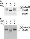
hRAD9 is phosphorylated in undamaged cells. (A) COS-1 cells transiently transfected with the construct expressing hRAD9m. After harvest, lysates were immunoprecipitated with α-myc 9E10 monoclonal antibody. Fractions of the immunoprecipitate were either treated or not with CIP in the presence or absence of orthovanadate (VO4). Samples were then subjected to Western analysis using antibody directed against the myc epitope. CIP treatment resulted in elimination of slower-migrating forms of hRAD9m, an effect that was not observed when VO4 was present. (B) Logarithmically growing HeLa cells were harvested and subjected to Western analysis using polyclonal α-hRAD9 antibodies. As in A, samples also treated with CIP in the presence or absence of vanadate, as indicated.
Figure 5
Figure 5
hRAD9 is located in the nucleus. Confocal immunofluorescence and light microscopy was performed on <50% confluent HeLa cells and 90% confluent HaCaT cells. Cells were fixed and probed with α-hRAD9 chicken polyclonal antibodies, followed by a fluorescently labeled anti-chicken IgY secondary antibody (A). DNA was visualized by staining with propidium iodide (B). Images from A and B were superimposed (C). The cellular borders of the HeLa and confluent HaCaT cells were visualized by light microscopy (D), and the light and fluorescent images were superimposed (E).

Similar articles

Cited by

References

    1. al-Khodairy F, Carr AM. DNA repair mutants defining G2 checkpoint pathways in Schizosaccharomyces pombe. EMBO J. 1992;11:1343–1350. - PMC - PubMed
    1. al-Khodairy F, Fotou E, Sheldrick KS, Griffiths DJ, Lehmann AR, Carr AM. Identification and characterization of new elements involved in checkpoint and feedback controls in fission yeast. Mol Biol Cell. 1994;5:147–160. - PMC - PubMed
    1. Altschul SF, Gish W, Miller W, Myers EW, Lipman DJ. Basic local alignment search tool. J Mol Biol. 1990;215:403–410. - PubMed
    1. Boddy MN, Furnari B, Mondesert O, Russell P. Replication checkpoint enforced by kinases Cds1 and Chk1. Science. 1998;280:909–912. - PubMed
    1. Cahill DP, Lengauer C, Yu J, Riggins GJ, Willson JK, Markowitz SD, Kinzler KW, Vogelstein B. Mutations of mitotic checkpoint genes in human cancers. Nature. 1998;392:300–303. - PubMed

Publication types

MeSH terms