. 1999 Jun;10(6):1997-2015.
doi: 10.1091/mbc.10.6.1997.

Vimentin dephosphorylation by protein phosphatase 2A is modulated by the targeting subunit B55

Affiliations
Free PMC article

Vimentin dephosphorylation by protein phosphatase 2A is modulated by the targeting subunit B55

P Turowski et al. Mol Biol Cell. 1999 Jun.
Free PMC article

Abstract

The intermediate filament protein vimentin is a major phosphoprotein in mammalian fibroblasts, and reversible phosphorylation plays a key role in its dynamic rearrangement. Selective inhibition of type 2A but not type 1 protein phosphatases led to hyperphosphorylation and concomitant disassembly of vimentin, characterized by a collapse into bundles around the nucleus. We have analyzed the potential role of one of the major protein phosphatase 2A (PP2A) regulatory subunits, B55, in vimentin dephosphorylation. In mammalian fibroblasts, B55 protein was distributed ubiquitously throughout the cytoplasm with a fraction associated to vimentin. Specific depletion of B55 in living cells by antisense B55 RNA was accompanied by disassembly and increased phosphorylation of vimentin, as when type 2A phosphatases were inhibited using okadaic acid. The presence of B55 was a prerequisite for PP2A to efficiently dephosphorylate vimentin in vitro or to induce filament reassembly in situ. Both biochemical fractionation and immunofluorescence analysis of detergent-extracted cells revealed that fractions of PP2Ac, PR65, and B55 were tightly associated with vimentin. Furthermore, vimentin-associated PP2A catalytic subunit was displaced in B55-depleted cells. Taken together these data show that, in mammalian fibroblasts, the intermediate filament protein vimentin is dephosphorylated by PP2A, an event targeted by B55.


Figures
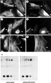
Figure 4
Figure 4
Vimentin disassembly and hyperphosphorylation after the functional knockout of B55. (A–D) Hs68 fibroblasts were microinjected with pECE-B55as and cultured for 6 h (A and B) or 12 h (C and D). Cells were then stained for vimentin (A and C). Arrowheads indicatemicroinjected cells as identified by costaining against the marker antibody (B and D). (E and F) Similarly, Hs68 fibroblasts were microinjected with a B56 antisense construct (manuscript in preparation) and subsequently stained for vimentin (E) and injection marker (F). Note that antisense B56 did not induce any changes in vimentin integrity. Bars, 5 μm. (G) Hs68 fibroblasts were grown on 1-mm2 glass chips and microinjected with the control plasmid pECE or the antisense B55-expressing construct pECE-B55as. Cells were subcultured for 6 h and then metabolically labeled with [32P]H3PO4 for 3 h. Subsequently, phosphoproteins were analyzed by two-dimensional gel electrophoresis as described in MATERIALS AND METHODS. Shown are portions of the autoradiograms containing vimentin (black arrowheads) and surrounding proteins only (white arrowheads indicate hsp 28). Shown are phosphoproteins of 230 cells injected with the control plasmid pECE (left panel) and 216 cells injected with the antisense B55 construct pECE-B55as (right panel). Autoradiographs were exposed for 4 d at −80°C and quantitated using a PhosphorImager.
Figure 1
Figure 1
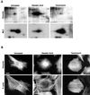
Analysis of phosphoproteins and the cytoskeleton of OA- and TAU-treated cells. (A) Equal numbers of Hs68 fibroblasts, grown on 12-mm glass coverslips, were treated with OA or TAU and metabolically labeled with [32P]H3PO4, and the totality of cellular proteins was separated by two-dimensional gel electrophoresis as described in MATERIALS AND METHODS. Shown are portions of the autoradiograms containing vimentin (upper panels) and myosin light chain (lower panels). Untreated, phosphoproteins of untreated cells labeled for 60 min; Okadaic Acid, phosphoproteins of cells treated with 1 μM OA during the 60-min labeling period; Tautomycin, phosphoproteins of cells treated with 10 μM TAU during 2 h and the subsequent 60-min labeling period. Autoradiographs were exposed at −80°C for 2 h for vimentin and for 6 h for MLC. (B) Hs68 fibroblasts were either left untreated or incubated with OA (1 μM for 45 min) or TAU (10 μM for 2 h). Subsequently they were fixed in formalin and costained for vimentin and F-actin using mAb V9 and Bodipy-phalloidin, respectively. Bar, 5 μm.
Figure 2
Figure 2
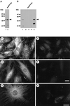
Subcellular distribution of the B55 subunit in Hs68 fibroblasts. (A) Approximately 20 μg of total cell extracts from Hs68 fibroblasts were electrophoretically separated (lane T) and immunoblotted; ∼10 ng of recombinant B55α (lane C) were included as positive control. Immunoblots were decorated with affinity-purified Ab55473/448. (B) Hs68 fibroblasts were differentially extracted for soluble cytoplasm (lane 1), insoluble cytoplasm (lane 2), nucleoplasm (lane 3), and the nuclear pellet also containing microfilament and IFs (lane 4). Twenty micrograms of the soluble (lanes 1 and 3) and equal volumes of the insoluble (lanes 2 and 4) fractions were electrophoretically separated and immunoblotted using Ab55recomb. An identical subcellular distribution was obtained using affinity-purified Ab55473/448. Note that both antibodies recognize different proteolytic fragments of recombinant B55 (lane C), namely an amino terminus with Ab55recomb and a carboxyl terminus for Ab55473/448. (C–H) Hs68 fibroblasts were grown on glass coverslips and formalin fixed. Cells were stained for B55 using either Ab55473/448 (C and D) or Ab55recomb (E–H). In D, F, and H, antibodies were preincubated with recombinant B55 as described in MATERIALS AND METHODS. The images resulting from the same antibody staining were acquired and treated identically. (G and H) Confocal section of ∼180 nm through the middle of the nucleus. Bar, 5 μm.
Figure 3
Figure 3
Depletion of cellular B55 by pECE-B55as. (A) Hs68 were transiently transfected with the empty pECE plasmid (mock) or with the B55 antisense construct pECE-B55as (B55as). Thirty-six hours after transfection 50 μg of total lysate were electrophoretically separated and immunoblotted using Ab55recomb. Immunodecoration using Ab55473/448 revealed a similar disappearance of B55 in extracts of pECE-B55as-transfected cells. In parallel experiments an efficiency of 60% transfection was determined using a green fluorescent protein expression construct. (B and C) Cells were microinjected with the B55-antisense RNA–expressing construct pECE-B55as, mixed with mouse immunoglobulin G, cultured for a further 15 h, fixed, and stained for B55 using Ab55473/448 (B). The arrowheads indicate the microinjected cell as identified by costaining for the microinjection marker antibody (C). Note that this cell has been exclusively microinjected in the nucleus. Bar, 5 μm.
Figure 6
Figure 6
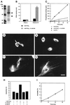
Enhanced dephosphorylation of vimentin in the presence of recombinant B55. (A) Twenty microliters of recombinant B55 prepared as described in MATERIALS AND METHODS were electrophoretically separated, and proteins were visualized using Coomassie brilliant blue. The molecular masses of marker proteins in kilodaltons (lane M) are indicated at the left. The major full-length and minor proteolytically degraded forms of B55, as identified by immunoblotting, are indicated with arrowheads. (B) The effect of B55 on the activity of purified dimeric PP2A2 toward phosphorylase and (p34cdc2 phosphorylated) histone H1 was assayed. Before the assay, PP2A2 (at 1 nM final concentration) was preincubated with (black bars) or without (hatched bars) ∼10 nM (final) recombinant B55 on ice. (C) Nitrocellulose strips, containing hyperphosphorylated [32P]vimentin were prepared as described in MATERIALS AND METHODS. Similarly to the phosphatase assays shown in B, strips containing equal amounts of [32P]vimentin (∼30,000 cpm and 5 μg of protein) were incubated with either 1 nM dimeric PP2A2 preincubated with (open squares) or without (closed squares) 10 nM recombinant B55 or with 1 nM catalytic subunit of PP1 (triangles). Phosphate release was determined and plotted against the assay time. (D–I) Hs68 fibroblasts grown on coverslips were treated with 1 μM OA for 1 h. Soluble proteins were extracted using Triton X-100 and 1.5 M KCl, and the remaining vimentin lattices were extensively washed in phosphatase assay buffer. Subsequently coverslips were overlaid with 30 μl of protein phosphatase assay buffer containing no phosphatase (D), 1 nM PP2A2 (E), or 1 nM PP2A2 and ∼10 nM recombinant B55 (F and G) and incubated at 37°C for 45 min. Reactions were stopped by formalin fixation and further stained for vimentin distribution. Bar, 5 μm. (H) Quantitative representation of the experiment shown in D–G; vimentin distribution resembling D or E was counted as bundled, whereas that resembling F or G or Figure 1B, upper left (unaffected vimentin), was counted as (re)assembled. On average, 600 stained vimentin lattices of each coverslip (i.e., assay point) were counted. In all assays ∼20% of the vimentin lattices had normal appearance and had not been affected by the initial OA treatment. (I) Time course of vimentin dephosphorylation by 1 nM PP2A2 and 10 nM B55. Vimentin reassembly was determined as in H, and for each time point the corresponding background (no PPase) was subtracted.
Figure 5
Figure 5
Microtubule and microfilament networks are unaffected in B55-depleted cells. Hs68 fibroblasts were microinjected with pECE-B55as and cultured for 12 h as in Figure 4. (A and B) Cells were then stained for tubulin (A). Arrowheads indicate microinjected cell as identified by costaining against the marker antibody (B). (C and D) Costaining for F-actin (C) and vimentin (D), respectively. Here the corresponding guinea pig marker antibody (revealed with amino-methylcoumarin) is not shown but indicated by arrowheads. In each case ∼50 cells were microinjected, and no effect on microtubule or actin distribution was observed. Bars, 5 μm.
Figure 7
Figure 7
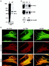
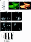
Association of PP2A with vimentin in Hs68 fibroblasts. (A and B) Vimentin was isolated from Hs68 fibroblasts by Triton X-100 and 0.6 M KCl extraction as described in MATERIALS AND METHODS. Material corresponding to one-third of a 100-mm dish of Hs68 was electrophoretically analyzed; equal proportions of the soluble (lanes S; S/10, formula image of S) and insoluble, vimentin-enriched (lanes VE) fractions were electrophoretically separated. Subsequently, proteins were stained with Coomassie brilliant blue (A). Arrows indicate the positions of vimentin (v) and actin (a). The migration of molecular mass markers is indicated at the left. Alternatively, gels were immunoblotted (B) for the presence of PP2Ac and PR65 using AbC302/309 (upper panel) and Ab65177/196 (middle panel). Approximately 5 ng of rabbit skeletal muscle PP2Ac and ∼10 ng of recombinant PR65 were included as a positive control (lanes C), and their positions are marked with arrowheads.Because of a high background staining in the region at ∼50 kDa, B55 could not be detected in an unequivocal way using our polyclonal Ab55. Vimentin was isolated from Hs68, transiently transfected to express HA-tagged B55. These preparations were immunoblotted using the monoclonal anti-HA (12CA5) (lower panel). nt, vimentin from nontransfected cells; t, from transfected cells. The arrowheads indicate the migration of HA-B55 as determined by anti-HA immunoprecipitates, run on the same gel and detected with Ab55recomb. (C) Hs68 fibroblasts were grown on glass coverslips and, before formalin fixation, extracted with 0.01% Triton X-100. Subsequently, cells were stained for PP2Ac, PR65, or B55 using AbCrecomb, Ab65177/196, or Ab55recomb, respectively, and costained for vimentin. The staining for PP2A subunits was revealed using FITC-conjugated reagents (upper panels), whereas vimentin was detected using Texas Red (middle panels). The lower panels show double exposures of the respective PP2A and vimentin staining (see MATERIALS AND METHODS). Bar, 5 μm.
Figure 8
Figure 8
Targeting by B55. (A) Immunoblot analysis of PP2A catalytic subunit from total cell lysates of Hs68 fibroblasts using AbC169/182 were performed similarly to those in Figure 1. Lane 1, ∼20 μg of cell extract; lane C, 5 ng of rabbit skeletal muscle PP2Ac. Positions of molecular mass markers are indicated at the left. The bands revealed are competed by the antigenic peptide. (B) Hs68 fibroblasts were grown on glass coverslips and formalin fixed. Subsequently, they were costained for the catalytic subunit using AbC169/182, revealed by FITC (left panel), and vimentin using mAb V9, revealed by Texas Red, as described for Figure 7B. To visualize colocalization of PP2Ac with vimentin, double exposures of both signal were recorded (right panel). Note that PP2Ac colocalized with most of the vimentin (yellow), but some regions of vimentin were devoid of PP2Ac staining (red, arrowheads). (C and D) Hs68 fibroblasts, grown on glass coverslips, were microinjected with the antisense B55 RNA–expressing construct pECE-B55as. After 12 h, cells were fixed using formalin. In C cells were stained for vimentin-associated PP2Ac using AbC169/182 (left panel) and for the microinjection marker (right panel). In D cells were stained stained for total PP2Ac using AbCrecomb (left panel) and for the microinjection marker (right panel). Arrowheads indicate injected cells. (E) In similar experiments as in C and D, cells were microinjected with the control plasmid pECE or with pECE-B55as. Subsequently they were stained for the totality of cellular PP2Ac using AbCrecomb (Turowski et al., 1995), vimentin-associated PP2Ac using AbC169/182, or vimentin using V9. Subsequently, the fluorescence of each microinjected cell was quantitated using ImgCalc. The histogram shows average fluorescence as determined from at least 30 injected cells for each staining. Values found for control-injected cells were arbitrarily set to 100%. Bars, 5 μm.

Similar articles

Cited by

References

    1. Agostinis P, Goris J, Waelkens E, Pinna LA, Marchiori F, Merlevede W. Dephosphorylation of phosphoproteins and synthetic phosphopeptides. Study of the specificity of the polycation-stimulated and Mg-ATP-dependent phosphorylase phosphatases. J Biol Chem. 1987;262:1060–1064. - PubMed
    1. Alessi DR, Andjelkovic M, Caudwell B, Cron P, Morrice N, Cohen P, Hemmings BA. Mechanism of activation of protein kinase B by insulin and IGF-1. EMBO J. 1996;15:6541–6551. - PMC - PubMed
    1. Andjelkovic N, Zolnierowicz S, Van-Hoof C, Goris J, Hemmings BA. The catalytic subunit of protein phosphatase 2A associates with the translation termination factor eRF1. EMBO J. 1996;15:7156–7167. - PMC - PubMed
    1. Blattler DP, Garner F, Van Slyke K, Bradley A. Quantitative electrophoresis in polyacrylamide gels of 2–40% J Chromatogr. 1972;64:147–155.
    1. Blose SH, Meltzer DI, Feramisco JR. 10-nm filaments are induced to collapse in living cells microinjected with monoclonal and polyclonal antibodies against tubulin. J Cell Biol. 1984;98:847–858. - PMC - PubMed

Publication types

MeSH terms

LinkOut - more resources