Comparative Study
. 1999 Jun;10(6):2063-74.
doi: 10.1091/mbc.10.6.2063.

Congruent docking of dimeric kinesin and ncd into three-dimensional electron cryomicroscopy maps of microtubule-motor ADP complexes

Affiliations
Free PMC article
Comparative Study

Congruent docking of dimeric kinesin and ncd into three-dimensional electron cryomicroscopy maps of microtubule-motor ADP complexes

K Hirose et al. Mol Biol Cell. 1999 Jun.
Free PMC article

Abstract

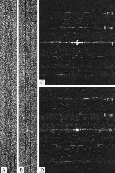
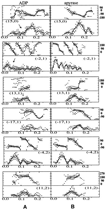
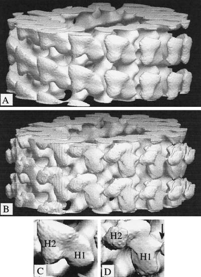
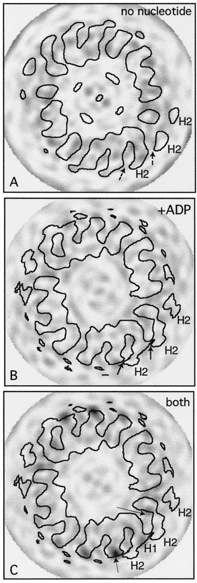
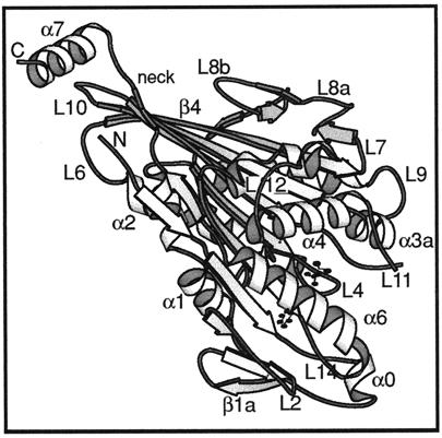
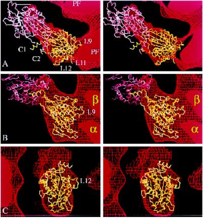
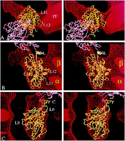
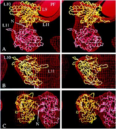
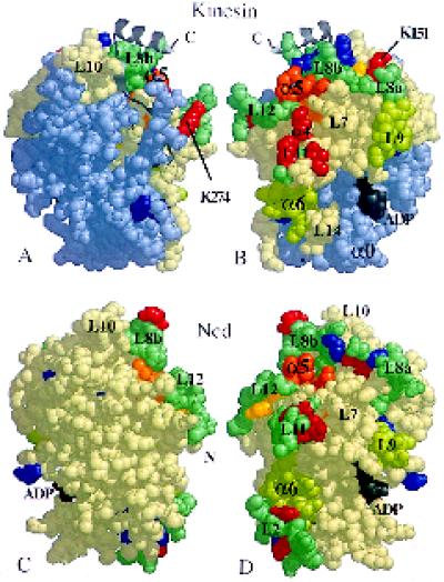
We present a new map showing dimeric kinesin bound to microtubules in the presence of ADP that was obtained by electron cryomicroscopy and image reconstruction. The directly bound monomer (first head) shows a different conformation from one in the more tightly bound empty state. This change in the first head is amplified as a movement of the second (tethered) head, which tilts upward. The atomic coordinates of kinesin.ADP dock into our map so that the tethered head associates with the bound head as in the kinesin dimer structure seen by x-ray crystallography. The new docking orientation avoids problems associated with previous predictions; it puts residues implicated by proteolysis-protection and mutagenesis studies near the microtubule but does not lead to steric interference between the coiled-coil tail and the microtubule surface. The observed conformational changes in the tightly bound states would probably bring some important residues closer to tubulin. As expected from the homology with kinesin, the atomic coordinates of nonclaret disjunctional protein (ncd).ADP dock in the same orientation into the attached head in a map of microtubules decorated with dimeric ncd.ADP. Our results support the idea that the observed direct interaction between the two heads is important at some stages of the mechanism by which kinesin moves processively along microtubules.


Figures

Figure 1
Figure 1
Electron microscope images and their diffraction patterns. (A and B) Images of 15-protofilament brain microtubules decorated with the double-headed kinesin motor construct (KΔ430) and either treated with apyrase to remove all free nucleotide (A) or in the presence of ADP and hexokinase (B). Bar, 50 nm. (C and D) Computed Fourier transforms from A and B. The equatorial (eq.) and 8-nm layer lines are relatively weaker in patterns from an ADP-containing complex (D) than in those from the nucleotide-free state (C).
Figure 2
Figure 2
Amplitudes and phases along the main layer lines of computed Fourier transforms. Phases (in degrees) are plotted in the upper half of each panel; amplitudes (all on the same scale) are shown in the bottom plots. The phases have been corrected for rotations about the microtubule axis and for shifts in the position of the phase origin along the axis to give the best overall agreement among different images. The data points are from individual images of 15-protofilament brain microtubules decorated with KΔ430 in ADP (A) or in the absence of nucleotides (effect of apyrase; B). The plotted lines show the averaged values. The layer-line indexing (n,l) refers to the Bessel order n, or number of equivalent helices in 15-protofilament microtubules, and the layer-line number l, based on a nominal 8-nm axial repeat. Some differences between nucleotide states are indicated by arrows: the phase distribution on the (15,0) layer line does not fall as steeply for ADP as for the tightly bound state; amplitudes on several layer lines are lower for the ADP state.
Figure 3
Figure 3
Surface representations of the 3-D density maps. (A and B) The maps were computed from averaged data (see Figure 2) for 15-protofilament brain microtubules decorated with dimeric kinesin in the absence of free nucleotide (A) or in the presence of ADP (B). They are in the standard orientation, with the microtubule plus end at the top of the page. (C and D) Enlarged views of individual dimeric motors are shown. In each case, H1 is the directly bound head; H2 is the tethered head. An arrow indicates the absence in the nucleotide-free structure (C) of a feature in the ADP-containing structure (D) that can fit loop L12 (see Figure 6c).
Figure 4
Figure 4
Contoured cross sections with variance levels. (A and B) Cross sections through the average maps in Figure 3 superimposed on variance values calculated from the individual maps. Arrows point to the projecting feature, sectioned at various levels, present in kinesin·ADP (B) but missing in the empty state (A). (C) Variancesobtained when nucleotide-free and ADP data sets are mixed. All variances are shown on the same scale, with darker density for higher values; there are peaks (arrows) on the exposed surface of the bound heads (H1), close to the point of attachment of the tethered heads (H2) and also close to where the tops of the bound heads contact tubulin, reflecting the changes described in the text.
Figure 5
Figure 5
Atomic structure of monomeric kinesin. A ribbon diagram of the kinesin motor domain (Sack et al., 1997) viewed from an orientation similar to that in a figure below (see Figure 6b). The heart-shaped catalytic domain comes to a point at the top left of the molecule. The neck (including β9 and β10) runs alongside leading to helix α7, part of the rod domain that forms a coiled coil in dimeric kinesin. Bound ADP shown as a ball and stick model. N and C, chain termini.
Figure 6
Figure 6
Fitting the atomic structure of kinesin into our kinesin·ADP map. Stereo views of two copies of the α-carbon backbone of the kinesin monomer (Sack et al., 1997) oriented inside part of the EM map (in Figure 3B) that shows tubulin protofilaments (PF) decorated with dimeric kinesin·ADP. (a) A top view, seen from the microtubule plus end. (b) A side view. (c) The view, at 90° to both a and b, showing the attached head from outside the microtubule. The map is represented as a surface net (red lines). One kinesin molecule (shown in yellow) occupies the attached head density; another (in pink) is positioned in the tethered head density. The relative orientation of the pink and yellow molecules mimics, as closely as possible, the asymmetric arrangement of heads in crystals of dimeric kinesin (Kozielski et al., 1997); thus, loop L10 of the attached head B contacts loop L8 of the tethered head A. A small distortion of each C-terminal helix (C1 and C2) would be needed to bring them together to form a coiled coil; this rod would then point almost directly away from the microtubule.
Figure 7
Figure 7
Alternative docking of kinesin into our kinesin·ADP map. (a–c) Stereo views showing two copies of the α-carbon backbone of monomeric kinesin oriented inside the same EM map of kinesin·ADP shown in Figure 6 but docked in the orientation favored by Hoenger et al. (1998). The directly bound head (yellow) fills the outer boundaries almost as well as in Figure 6, but the top (a) and side (c) views fit less accurately, and in the side view (b), the complementarity of kinesin’s inside surface with the tubulin subunits (α and β) is less good. A second molecule (pink) arranged as in the crystallographic structure is completely out of the EM density, as can be seen in a. The coiled coil would point obliquely into the microtubule surface. C, C terminus; PF, protofilament.
Figure 8
Figure 8
Fitting the atomic structure of ncd into our ncd·ADP map. (a–c) Stereo views of the α-carbon backbone of monomeric ncd (Sablin et al., 1996) oriented inside a map showing tubulin protofilaments (PF) decorated with dimeric ncd with bound ADP (Hirose et al., 1998). As in Figures 6 and 7, the map is represented at a single contour level as a surface net (red lines). One copy of the monomeric ncd crystal structure (yellow) is positioned in the attached head density; another (pink) lies in the tethered head density. The labeled loops (L11, L9, etc.) are equivalent to those in the kinesin structure (see Figure 5). The side view (b) at 90° to a is shown with the tethered head removed. The front view in c is at 90° to both a and b. There is an approximate twofold axis between the heads, running almost vertically in c. N, N terminus.
Figure 9
Figure 9
Exposed and protected surfaces of the bound motor domains. Space-filling images of monomeric kinesin and Ncd, showing the positions of residues that appear to be important. (A and C) The views from directly outside a decorated microtubule for heads bound in the orientation shown in Figure 6; the head (B and D) has been rotated by 180° to show the surface closest to tubulin. Cleavable residues are either protected from proteolysis by microtubules (red), including lysines K151 and K274, or always exposed (blue) (Alonso et al., 1998); yellow residues are cut differentially in ADP versus AMP·PNP, in the absence of microtubules. Alanine scanning (Woehlke et al., 1997) of kinesin suggests that the orange arginine and lysine residues (R280, K283, and R286—corresponding to R278, K281, and R284 in human kinesin, respectively) in the L12/α5 region are important for the binding of microtubules. Some predicted tubulin-binding loops (L7, L8, L11, and L12 [Kull et al., 1996]) are shown in blue-green. Helices α4 and α6 and loops L7 and L9 also appear to be exposed to tubulin. The light-blue part of kinesin represents residues 1–130 (including α0), which can be removed without loss of microtubule binding (Yang et al., 1989). N and C, chain termini.

Similar articles

Cited by

References

    1. Alonso MC, Vanderkerckhove J, Cross RA. Proteolytic mapping of kinesin/ncd-microtubule interface: nucleotide-dependent conformational changes in the loops L8 and L12. EMBO J. 1998;17:945–951. - PMC - PubMed
    1. Amos LA, Hirose K. The structure of microtubule-motor complexes. Curr Opin Cell Biol. 1997;9:4–11. - PubMed
    1. Arnal I, Metoz F, DeBonis S, Wade RH. Three-dimensional structure of functional motor proteins on microtubules. Curr Biol. 1996;6:1265–1270. - PubMed
    1. Arnal I, Wade RH. Nucleotide-dependent conformations of the kinesin dimer interacting with microtubules. Structure. 1998;6:33–38. - PubMed
    1. Crevel IM-T, Lockhart A, Cross RA. Weak and strong states of kinesin and ncd. J Mol Biol. 1996;257:66–76. - PubMed

Publication types

LinkOut - more resources