. 1999 Jun;10(6):2075-86.
doi: 10.1091/mbc.10.6.2075.

Activation of utrophin promoter by heregulin via the ets-related transcription factor complex GA-binding protein alpha/beta

Affiliations
Free PMC article

Activation of utrophin promoter by heregulin via the ets-related transcription factor complex GA-binding protein alpha/beta

T S Khurana et al. Mol Biol Cell. 1999 Jun.
Free PMC article

Abstract

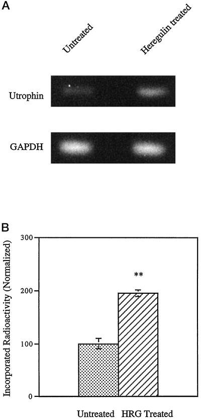
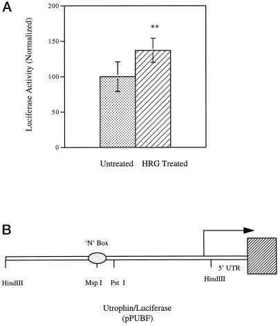
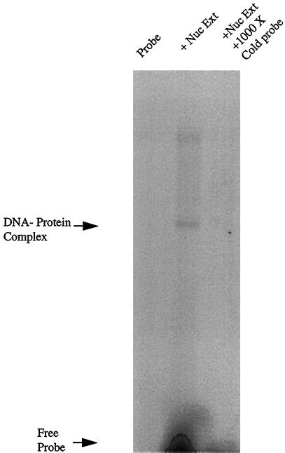
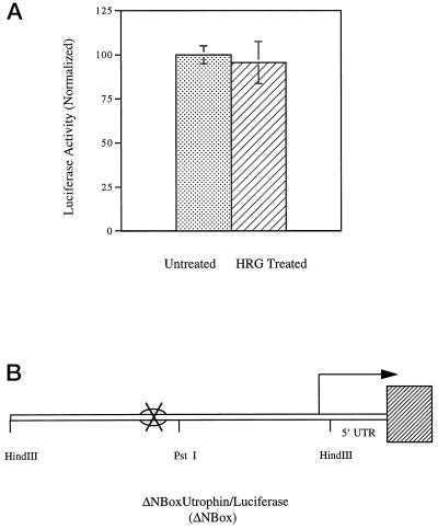
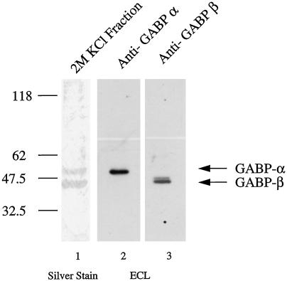
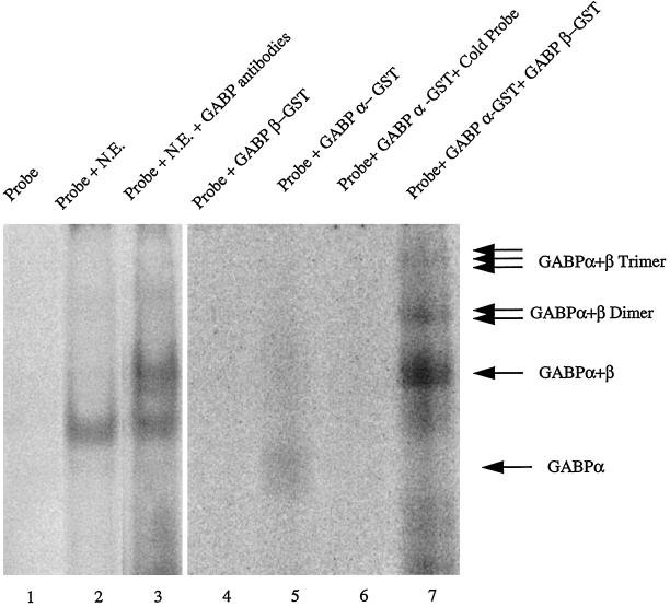
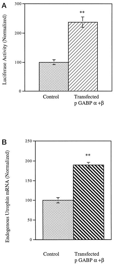
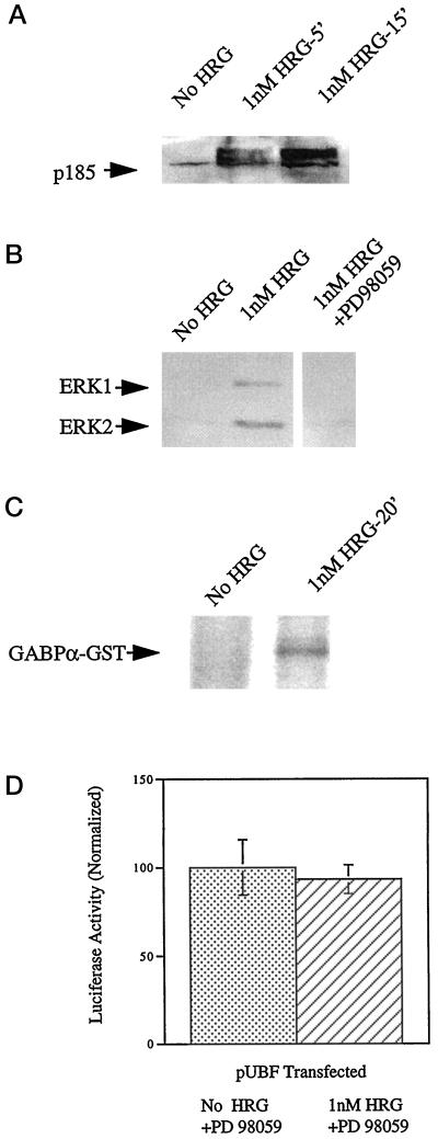
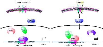
Utrophin/dystrophin-related protein is the autosomal homologue of the chromosome X-encoded dystrophin protein. In adult skeletal muscle, utrophin is highly enriched at the neuromuscular junction. However, the molecular mechanisms underlying regulation of utrophin gene expression are yet to be defined. Here we demonstrate that the growth factor heregulin increases de novo utrophin transcription in muscle cell cultures. Using mutant reporter constructs of the utrophin promoter, we define the N-box region of the promoter as critical for heregulin-mediated activation. Using this region of the utrophin promoter for DNA affinity purification, immunoblots, in vitro kinase assays, electrophoretic mobility shift assays, and in vitro expression in cultured muscle cells, we demonstrate that ets-related GA-binding protein alpha/beta transcription factors are activators of the utrophin promoter. Taken together, these results suggest that the GA-binding protein alpha/beta complex of transcription factors binds and activates the utrophin promoter in response to heregulin-activated extracellular signal-regulated kinase in muscle cell cultures. These findings suggest methods for achieving utrophin up-regulation in Duchenne's muscular dystrophy as well as mechanisms by which neurite-derived growth factors such as heregulin may influence the regulation of utrophin gene expression and subsequent enrichment at the neuromuscular junction of skeletal muscle.


Figures

Figure 1
Figure 1
Heregulin increases utrophin mRNA in skeletal muscle cultures. Differentiated L6 rat myotubes were incubated with 1 nM heregulin in PBS for 30 min along with controls. RNA was extracted, and quantitative RT-PCR performed. (A) Representative experiment showing the 322-bp utrophin fragment and the 194-bp GAPDH control fragment obtained by RT-PCR. (B) Results of radioactive quantification of four individual experiments taken together. The stippled bars represent utrophin mRNA levels in untreated cells, and cross-hatched bars represent the levels in heregulin (HRG)-treated cultures. Heregulin treatment increases the endogenous utrophin message in muscle cell cultures to 195% of control levels. Error bars indicate SEM; n = 4). Asterisks denote the results were statistically highly significant at p < 0.001.
Figure 2
Figure 2
Heregulin activates the utrophin promoter in muscle cell cultures. The utrophin promoter–luciferase reporter construct pPUBF was cotransfected into L6 muscle cell lines or mouse primary muscle cultures along with transfection control plasmid pRL and assayed after 24–48 h of incubation with heregulin-containing medium or control-containing medium. (A) pPUBF derived firefly luciferase activity is normalized to pRL-derived renilla luciferase activity (internal control) as 100% in the untreated group and expressed as luciferase activity (normalized). The graph represents the summary of 10 individual experiments, 5 sets of experiments in primary mouse cultures and 5 sets of experiments in L6 rat muscle cell cultures. The stippled bars represent utrophin promoter activity in untreated cells, and cross-hatched bars represent the levels in heregulin (HRG)-treated cultures. (B) Schematic of the pPUBF-luciferase construct (Dennis et al., 1996) and was kind gift from Drs. J. Tinsley and K. Davies. Heregulin increases de novo utrophin transcription in muscle cell cultures to 138% of control levels. Error bars indicate SEM; n = 60. Asterisks denote the results were statistically highly significant at p < 0.001.
Figure 3
Figure 3
The utrophin N-Box binds proteins in nuclear extracts of cultured L6 myotubes. EMSA was performed with the radiolabeled UtroNBox probe using 10 μg of nuclear extract made from cultured L6 myotubes. Lane 1, migration of free, unretarded probe control; lane 2, mobility shift of the probe in the presence of a N-box–binding factor present in L6 nuclear extracts; lane 3, control for specificity showing that the mobility shift was competed with a 1000× excess of unlabeled probe.
Figure 4
Figure 4
The N-Box motif in the utrophin promoter mediates transcriptional activation by heregulin in cultured muscle cells. The mutant utrophin promoter–luciferase reporter construct ΔNBox (deleted in the N-box) was cotransfected into L6 muscle cell lines along with transfection control plasmid pRL and assayed after 24 h of incubation with either heregulin-containing medium or controls. (A) The ΔNBox-derived firefly luciferase activity is normalized to pRL-derived renilla luciferase activity (internal control) as 100% in the untreated state and expressed as luciferase activity (normalized). The stippled bars represent mutant utrophin promoter activity in untreated cells, and cross-hatched bars indicate the levels in heregulin (HRG)-treated cultures. (B) Schematic of the ΔNBox–luciferase construct; the cross depicts the site of deletion mutation removing the N-box. Heregulin does not activate the mutant utrophin promoter bearing a deletion of the N-Box. Error bars indicate SEM; n = 36. The differences of expression are not statistically significant.
Figure 5
Figure 5
Identification of transcription factors that bind the N-Box of the utrophin promoter. UtroNBox-coupled magnetic particles were used to perform DNA affinity chromatography to purify promoter-binding proteins from nuclear extracts of cultured L6 myotubes. Fifty micrograms of nuclear extracts were used, and binding proteins were eluted in 25 μl of 2 M KCl. A 15-μl aliquot was resolved using 12% SDS-PAGE gels and subjected to silver staining (lane 1), or 5-μl aliquots were immunoblotted with affinity-purified anti-GABPα and -GABPβ antibodies and subjected to enhanced chemiluminescence detection (lanes 2 and 3). Lane 1, silver-stained proteins demonstrating that the molecular masses of purified proteins (43 and 58 kDa) exactly matches the molecular masses of GABPβ and GABPα heterodimeric complex of transcription factors; lane 2, the 58-kDa band in the purified fraction is recognized by affinity-purified anti-GABPα antibodies; lane 3, the 43-kDa band in the purified fraction is recognized by affinity-purified anti-GABPβ antibodies. The additional high-molecular-mass species presumably represents a GABPβ isoform sharing sequence similarity with the GABPβ1 isoform, against which the antibodies were raised. The anti-GABPβ1 antibodies used in this study (see MATERIALS AND METHODS) are predicted to recognize all GABPβ isoforms.
Figure 6
Figure 6
The utrophin N-Box binds the heterodimeric GABPα/β transcription factor. EMSA was performed with the radiolabeled oligonucleotide UtroNBox probe, nuclear extracts, and antibodies against GABPα/β (for supershift assays) in lanes 1–3. In lanes 4–7 EMSA was performed using purified GABPα and GABPβ GST fusion proteins and this probe. Lane 1, lack of mobility shift when the probe is electrophoresed alone; lane 2, mobility shift when the probe is incubated with nuclear extracts from cultured muscle cells; lane 3, the addition of antibodies directed against GABPα/β causes a further mobility shift or supershift compared with the mobility shift obtained using nuclear extracts alone; lane 4, lack of mobility shift using the GABPβ protein, suggesting that the UtroNBox probe does not bind GABPβ by itself; lane 5, mobility shift when the probe is incubated with GABPα fusion protein; lane 6, specificity of the interaction with GABPα, because it is competed with a 1000× excess of unlabeled probe; lane 7, mobility shifts with the formation of GABPα/β multimers when the probe is incubated with both GABPα and GABPβ fusion proteins, suggesting enhancement of GABPα binding by reconstitution of the heterodimeric GABPα/β transcription factor complex.
Figure 7
Figure 7
GABPα/β activates the utrophin promoter in muscle cell cultures. (A) The utrophin promoter–luciferase reporter construct PUBF was cotransfected into L6 muscle cell lines along with expression constructs pGABPα, pGABPβ, and pCAGGS (empty vector) along with transfection control pRL and assayed after 24 h of incubation. PUBF-derived firefly luciferase activity is normalized to pRL-derived renilla luciferase activity (internal control) in control transfectants as 100% and expressed as luciferase activity (normalized). The stippled bars represent utrophin promoter activity in cells transfected with empty vector pCAGGS, and cross-hatched bars represent the levels in cultures transfected with pGABPα and pGABPβ. (B) L6 muscle cell lines were transfected with expression constructs pGABPα, pGABPβ, and pCAGGS (empty vector) as control. RNA was extracted, and quantitative RT-PCR was performed for estimating the level of utrophin mRNA. The graph shows the results of quantification of five individual experiments. The stippled bars represent utrophin mRNA levels in control cultures, and cross-hatched bars represent the levels in cultures transfected withpGABPα and pGABPβ. GABPα and GABPβ cotransfection increases de novo utrophin transcription in muscle cell cultures to 238% of control levels (error bars indicate SEM; n = 24) and increases endogenous utrophin mRNA by 189% of control levels (error bars indicate SD; n = 5). Asterisks denote the results were statistically highly significant at p < 0.001.
Figure 8
Figure 8
Heregulin-mediated utrophin promoter activation requires ERK activation. (A) Differentiated L6 rat myotubes were incubated with PBS alone or 1 nM heregulin for 5 and 15 min. Proteins were extracted from cultures and resolved on 10% SDS-polyacrylamide gels and electroblotted onto polyvinylidene difluoride membranes followed by Western blotting with the PY20 antibody to detect phospotyrosine residues. The blot shows a mobility shift and increased tyrosine phosphorylation (activation) of p185 (HER receptor) species and serves as a positive control. (B) Proteins were extracted from control cultures, cultures treated for 20 min with 1 nM HRG, and cultures preincubated with PD98059 before treatment with HRG. Proteins were resolved on 10% SDS-polyacrylamide gels and electroblotted onto polyvinylidene difluoride membranes followed by Western blotting using the pTEpY antibody, which detects dually phosphorylated, active forms of ERK. The blot shows activation of ERK1 and ERK2 by heregulin and prevention of activation by preincubation with PD 98059. (C) In vitro kinase assay was performed using immunoprecipitated ERK from control or muscle cell cultures treated with 1 nM heregulin for 20 min. Proteins were resolved on 10% SDS-polyacrylamide gels, and radioactively labeled proteins were detected by autoradiography. The autoradiograph shows that activated ERK extracted from heregulin-treated cells could phosphorylate the GST-GABPα fusion protein. (D) The utrophin promoter–luciferase reporter construct pPUBF was cotransfected into L6 muscle cell lines along with transfection control plasmid pRL and assayed after 48 h of incubation with PD98059 in heregulin-containing medium or control medium. The stippled bars represent normalized utrophin promoter (luciferase) activity in untreated cells, and cross-hatched bars represent the levels in heregulin-treated cultures. Heregulin does not activate the utrophin promoter in muscle cell cultures treated with PD98059. Error bars indicate SD; n = 6. The differences of expression are not statistically significant.
Figure 9
Figure 9
Model for utrophin up-regulation by GABPα/β in muscle cell cultures. In this schematic we propose that in unstimulated muscle cultures (and adult muscle) utrophin is transcribed at low levels, possibly because of transcriptional repression activity at the ets-binding site by repressors such as ERF or ERF-like molecules. Upon stimulation with heregulin, transcription is activated via the MAP kinase pathways by decreasing the repressor activity, as well as increasing the propensity of GABPα/β transcription factors to bind and heterodimerize, leading to an overall increase of utrophin transcription. This sequence is from human utrophin promoter and shows the relative position, content, and overlap of the N-box (turquoise box) and the site bound by the ets transcription factor complex GABPα/β (lilac box) in muscle cell cultures to activate the transcription of utrophin.

Similar articles

Cited by

References

    1. Ausubel F, et al. Current Protocols in Molecular Biology. New York: Wiley; 1995.
    1. Brown RH. Dystrophin-associated proteins and muscular dystrophies. Annu Rev Med. 1997;48:457–466. - PubMed
    1. Brown TA, McKnight SL. Specificities of protein-protein and protein-DNA interaction of GABP alpha and two newly defined ets-related proteins. Genes & Dev. 1992;6:2502–2512. - PubMed
    1. Campbell KP. Three muscular dystrophies: loss of cytoskeletal extracellular matrix linkage. Cell. 1996;80:675–679. - PubMed
    1. Campbell KP, Crosbie RH. Muscular dystrophy. Utrophin to the rescue. Nature. 1996;384:308–309. - PubMed

Publication types

MeSH terms

LinkOut - more resources