

Interfacial membrane docking of cytosolic phospholipase A2 C2 domain using electrostatic potential-modulated spin relaxation magnetic resonance
- PMID: 10359764
- PMCID: PMC21967
- DOI: 10.1073/pnas.96.12.6637
Interfacial membrane docking of cytosolic phospholipase A2 C2 domain using electrostatic potential-modulated spin relaxation magnetic resonance
Abstract
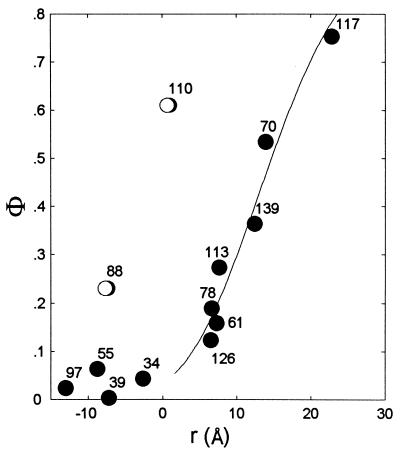
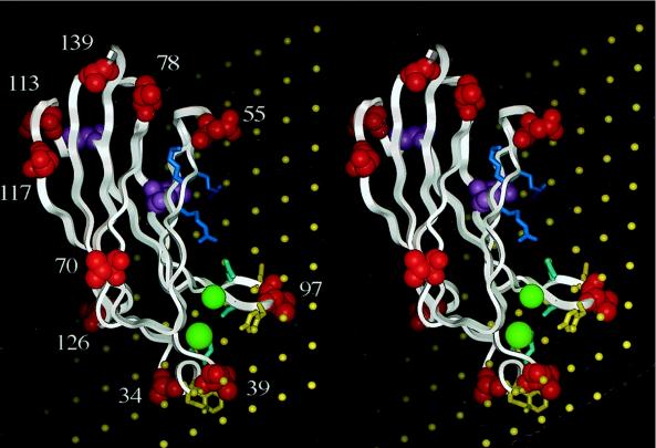
The C2 domain of cytosolic phospholipase A2 (C2cPLA2) plays an important role in calcium-dependent transfer of the protein from the cytosol to internal cellular membranes as a prelude for arachidonate release from membrane phospholipids. By using a recently developed electron paramagnetic resonance approach together with 13 site-specifically nitroxide spin labeled C2cPLA2s and membrane-permeant and -impermeant spin relaxants, we have determined the orientation of C2cPLA2 with respect to the surface of vesicles of the phospholipid 1,2-dioleoyl-sn-glycero-3-phosphomethanol. The structure reveals that the two calcium-binding regions on C2cPLA2 that display hydrophobic residues, CBR1 and CBR3, are partially inserted into the core of the membrane. CBR2 that contains predominantly hydrophilic residues is close to the membrane but not inserted. The long axis of the cylindrical C2cPLA2 molecule is tilted with respect to the bilayer normal, which brings a cluster of basic protein residues close to the phospholipid headgroups. Such an orientation places the two bound calcium ions close to the membrane surface. All together, the results provide structural support for previous proposals that binding of C2cPLA2 to the membrane interface is driven in part by insertion of hydrophobic surface loops into the membrane core. The results are contrasted with previous studies of the interfacial binding of the first C2 domain of synaptotagmin I, which has shorter surface loops that display basic residues for electrostatic interaction with the bilayer surface.
Figures
References
-
- Wymann M P, Pirola L. Biochim Biophys Acta. 1998;1436:127–150. - PubMed
-
- Plant P J, Yeger H, Staub O, Howard P, Rotin D. J Biol Chem. 1997;297:32329–32336. - PubMed
-
- Ibata K, Fukuda M, Mikoshiba K. J Biol Chem. 1998;273:12267–12273. - PubMed
-
- Rizo J, Sudhof T C. J Biol Chem. 1998;273:15879–15882. - PubMed
MeSH terms
Substances
LinkOut - more resources
Full Text Sources
Miscellaneous
