. 1999 Jun 8;96(12):6728-33.
doi: 10.1073/pnas.96.12.6728.

Signaling via beta1 integrins and mitogen-activated protein kinase determines human epidermal stem cell fate in vitro

Affiliations

Signaling via beta1 integrins and mitogen-activated protein kinase determines human epidermal stem cell fate in vitro

A J Zhu et al. Proc Natl Acad Sci U S A. .

Abstract

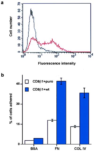
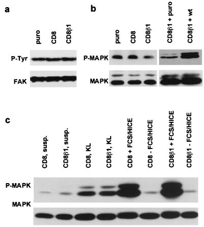
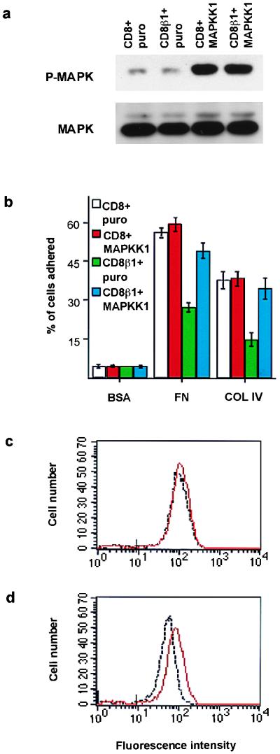
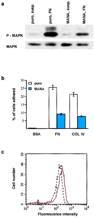
Human epidermal stem cells express higher levels of beta1 integrins and are more adhesive than keratinocytes that are destined to differentiate. To investigate whether high beta1 integrin expression and adhesiveness are essential for maintaining keratinocytes in the stem cell compartment, we introduced a dominant-negative beta1 integrin mutant, CD8beta1, into cultured human keratinocytes, thereby interfering with beta1 integrin function. Surface beta1 integrin levels, adhesiveness, and mitogen-activated protein (MAP) kinase activation on fibronectin were reduced, and exit from the stem cell compartment was stimulated. Adhesiveness and proliferative potential were restored by overexpressing wild-type beta1 integrin or by constitutive MAP kinase activation. Conversely, a dominant-negative MAP kinase kinase 1 mutant decreased adhesiveness and stem cell number in the absence of CD8beta1. MAP kinase activation by alpha6beta4-mediated adhesion and mitogens was normal in CD8beta1 cells, and constitutive MAP kinase activation did not affect adhesion and proliferation of control keratinocytes. We conclude that beta1 integrins and MAP kinase cooperate to maintain the epidermal stem cell compartment in vitro.


Figures

Figure 1
Figure 1
Subpopulations of human epidermal keratinocytes. Each subpopulation can be distinguished on the basis of β1 integrin expression or function (2, 4, 6).
Figure 2
Figure 2
Effect of CD8β1 on keratinocyte adhesiveness. (a and b) Cell-surface CD8 levels were evaluated by flow cytometry in puro- (black), CD8- (red in a), and CD8β1- (red in b) expressing keratinocytes. Profiles show total population. (c) Cells (104 per well) were plated for 3 hr in the presence of 25 μM cycloheximide. (d) Equal numbers of cells (103 per 35 mm dish) expressing puro, CD8, or CD8β1 were plated overnight on tissue culture plastic (TCP), type IV collagen (COL IV, 50 μg/ml), fibronectin (FN, 25 μg/ml) or keratinocyte extracellular matrix enriched for laminin 5 (KL) in the presence of J2–3T3 feeder cells, and the number of adherent keratinocytes was determined. (Error bars in c and d = standard deviation of the mean of triplicate samples within one experiment.)
Figure 3
Figure 3
Effects of CD8β1 on growth and terminal differentiation. (a) 500 (CD8, puro) or 650 (CD8β1) cells were plated per 35 mm dish, and triplicate dishes were harvested on the days shown. (b) The proportion of involucrin-positive keratinocytes was determined in preconfluent adherent cultures (before suspension) and after suspension for 24 hr in methyl cellulose-supplemented medium. [Error bars = standard deviation of the mean of triplicate (a) and duplicate (b) samples within one experiment.]
Figure 4
Figure 4
Effects of CD8β1 on β1 integrin expression and clonogenicity. (a and b) Flow cytometry of basal keratinocytes with an antibody to the β1 integrin extracellular domain, CD29-FITC. Black lines: puro-expressing cells; red lines: CD8- (a) or CD8β1- (b) expressing cells. Marks on x axes: maximum fluorescence of cells labeled with K9–18-FITC. (c) 500 (CD8, puro) or 650 (CD8β1) cells were plated on tissue culture plastic- (TCP), 50 μg/ml type IV collagen- (COL IV), or 25 μg/ml fibronectin- (FN) coated 35-mm dishes and cultured for 14 days; rhodanile blue staining.
Figure 5
Figure 5
Introduction of wild-type chicken β1 integrin subunit (wt) into keratinocytes expressing CD8β1. (a) Flow cytometry of basal keratinocytes with JG22 to chicken β1 integrin. Black line: cells expressing CD8β1 and puro. Red line: cells expressing CD8β1 and chicken β1 integrin (CD8β1 + wt). (b) Doubly infected cells (104 ) were plated for 3 hr with 25 μM cycloheximide on heat-denatured BSA (0.5 mg/ml), fibronectin (FN, 25 μg/ml), or type IV collagen (COL IV, 50 μg/ml). (Error bars = standard deviation of the mean of triplicate samples in one experiment.)
Figure 6
Figure 6
FAK and MAPK phosphorylation in CD8-, CD8β1-, and CD8β1 + wild type-expressing cells. (a—c) Western blots showing tyrosine phosphorylation of FAK (P-Tyr) (a) and threonine/tyrosine phosphorylation of MAPK (P-MAPK). (b and c Lower) Loading controls for FAK (a) and MAPK (p42mapk and p44mapk) (b and c). (a and b) Cells were plated on fibronectin for 30 min in serum-free medium. (c) Cells were held in suspension for 30 min (susp.), plated on keratinocyte ECM for 30 min in serum-free medium (KL), or plated on 25 μg/ml fibronectin in serum-free medium for 4 hr and then treated with fresh serum-free medium (−FCS/HICE) or complete medium (+FCS/HICE) for 15 min.
Figure 7
Figure 7
Effects of MAPKK1 on adhesion and integrin expression. (a) Western blot showing threonine/tyrosine phosphorylation of MAPK (P-MAPK). (Lower) Loading control (p42mapk and p44mapk). (b) Adhesion assay performed as in Fig. 5 b. (c and d) Flow cytometry analysis of surface β1 integrin levels on basal keratinocytes. Black lines: CD8- (c) or CD8β1- (d) expressing cells infected with puro. Red lines: CD8- (c) or CD8β1- (d) expressing cells infected with activated MAPKK1. Marks on x axes: maximum fluorescence of cells labeled with K9–18-FITC.
Figure 8
Figure 8
Effects of a dominant-negative MAPKK1 mutation (MANA) adhesion and integrin expression. (a) Western blot showing threonine/tyrosine phosphorylation of MAPK (P-MAPK) in cells plated on 25 μg/ml fibronectin (FN) or held in suspension (susp.) for 30 min. (b) Adhesion assay performed as in Fig. 5b. (c) Flow cytometry analysis of surface β1 integrin levels on basal keratinocytes. Red line: puro-expressing cells; black line: MANA-expressing cells. Mark on x axis: maximum fluorescence of cells labeled with UCHT4-FITC.

Similar articles

Cited by

References

    1. Hall P A, Watt F M. Development (Cambridge, UK) 1989;106:619–633. - PubMed
    1. Jones P H, Watt F M. Cell. 1993;73:713–724. - PubMed
    1. Watt F M. Philos Trans R Soc London B. 1998;353:831–837. - PMC - PubMed
    1. Jones P H, Harper S, Watt F M. Cell. 1995;80:83–93. - PubMed
    1. Gandarillas A, Watt F M. Genes Dev. 1997;11:2869–2882. - PMC - PubMed

Publication types

Substances