. 1999 Jun 8;96(12):6757-62.
doi: 10.1073/pnas.96.12.6757.

Control of the low voltage-activated calcium channel of mouse sperm by egg ZP3 and by membrane hyperpolarization during capacitation

Affiliations

Control of the low voltage-activated calcium channel of mouse sperm by egg ZP3 and by membrane hyperpolarization during capacitation

C Arnoult et al. Proc Natl Acad Sci U S A. .

Abstract

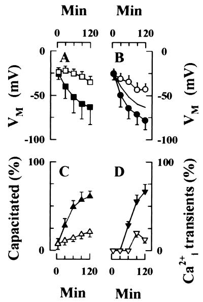
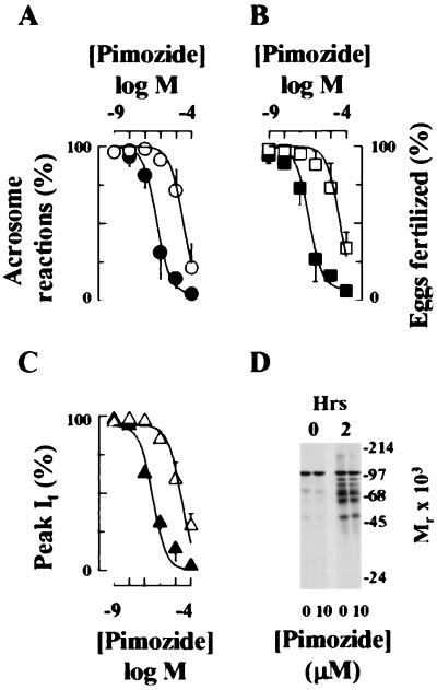
Sperm adhesion to egg zonae pellucidae initiates sperm acrosome reactions, an exocytotic event that is an early step during fertilization. Previously, it was suggested that zona pellucida-evoked Ca2+ entry into sperm through low voltage-activated Ca2+ channels is an essential step in acrosome reactions, based on the inhibitory effects of Ca2+ channel antagonists. However, analysis of this channel is limited by the inability to apply electrophysiological methods directly to sperm. In this report, optical methods of determining membrane potential and internal Ca2+ levels were used to demonstrate that (i) contact with zonae pellucidae activates a transient Ca2+ response in sperm that has a time course and antagonist sensitivity anticipated of low voltage-activated Ca2+ channels; (ii) these channels are unavailable for opening in uncapacitated sperm because of voltage-dependent, steady state inactivation; (iii) membrane hyperpolarization during sperm capacitation is sufficient to recruit channels into a closed state, from which they are available for opening during fertilization; and (iv) channel conductance state may be a factor in determines the efficacy with which channel antagonists inhibit fertilization. This study provides evidence for the activation of sperm Ca2+ channels during gamete adhesion and offers a mechanism that may account for aspects of the regulation of sperm fertility during capacitation through the control of channel availability. Finally, these results suggest that channel conductance state may be a central feature in the design of channel antagonists that inhibit sperm function.


Figures

Figure 1
Figure 1
ZP3 produces Ca2+i transients that are detected with mag-fura2. (A) Dye (λEX, 360 nm; λEM, 510 nm) distributes throughout sperm and permits ratiometric image acquisition from all regions. (B) Ca2+i responses to ZP3 treatment in the absence (○) and the presence (●) of PN200–110. Cells were treated with buffer (b) or with 1 μM PN200–110 (pn) for 10 min before addition of ZP3 (10 μg/ml; bar). The rising phase of the ZP3-dependent Ca2+i response in the absence of channel antagonists is approximated by the expression Ca2+i = et, where t and τ represent the time and the time constant, respectively (—). In this cell, τ = 12.7 msec. The initial portion of the relaxation trace is approximated by the expression Ca2+i = ae−t/τ, where a designates the maximal response (- - -) and where a = 9.11 μM and τ = 40.9 msec. (C) Peak Ca2+i responses to ZP (40 μg/ml) or to ZP3 (10 μg/ml) are inhibited by La3+ (○) and by Cd2+ (●). Data points represent the means (±SD) of observations on 4–7 different cells and are approximated by the expression R = (100 ∗ IC50)/(IC50 + D). R is observed response; D is antagonist concentration; and IC50 is the drug concentration producing 50% inhibition. Derived IC50 values were La3+, 171 ± 63 μM; and Cd2+, 652 ± 134 μM. Results using ZP3 and ZP are indistinguishable and are pooled for presentation. (D) ZP3-dependent Ca2+i transients are inhibited by LVA current antagonists. Pimozide was added at 15 min of a 120-min incubation in a capacitating medium whereas PN200–110 and Ni2+ were added at 90 min. After incubation for 120 min, sperm were treated with ZP3. Data represent peak Ca2+i values (mean ± SD). The fraction of cells in each treatment group that exhibit transients is indicated. Total cells were: buffer, 12; fetuin (40 μg/ml), 14; ZP (40 μg/ml), 26; ZP2 (40 μg/ml), 14; ZP3 (10 μg/ml), 37; +PN200–110 (0.1 μM), 9; +PN200–110 (1 μM), 11; +pimozide (0.1 μM), 12; +pimozide (1 μM), 16; +Ni2+ (5 μM), 8; +Ni2+ (50 μM), 7.
Figure 2
Figure 2
Capacitation results in membrane hyperpolarization. (A) Hyperpolarization of sperm populations during capacitation. VM values of immobilized sperm were calculated from di8-ANEPPS emission of single cells during a 120-min incubation under conditions that do (■, 75 cells) or do not support capacitation (□, 61 cells). Data are the mean (±SD) of 5–8 different experiments and entail determinations from the indicated number of cells. (B) Membrane hyperpolarization is associated with capacitation. Capacitation state of individual sperm was determined by ability of sperm to initiate exocytosis during a subsequent challenge with ZP. Data from 75 cells were summed to assemble population VM values (—; redrawn from A), as well as from the subpopulations that are (●) or are not (○) capacitated by 120 min. Strong hyperpolarization is restricted to the subpopulation of sperm that capacitate. (C) The time course of capacitation was determined by CTC fluorescence emission. Motile sperm were incubated in a capacitating medium (▴) or in a medium that does not capacitate mouse sperm efficiently (▵). Data are the mean ± SD of triplicate experiments, with 200 sperm assayed/time point in each experiment. (D) Sperm develop the ability to generate ZP/ZP3-dependent Ca2+i transients during capacitation. Sperm were incubated for the indicated times either in a capacitating medium (▾) or in a noncapacitating medium (▿), as described in C and were stimulated with either ZP (40 μg/ml) or ZP3 (10 μg/ml), and Ca2+i responses were recorded. Data represents mean (±SD) of observations on 3–8 cells, with ZP- and ZP3-stimulated responses pooled for presentation.
Figure 3
Figure 3
Effects of capacitation state on the inhibition of ZP3-dependent exocytosis and fertilization by Ca2+ channel antagonists. (A and B) Pimozide was added to sperm either at 15 min (●, ■) or at 90 min (○, □) of a 120-min incubation in a capacitating medium. Sperm subsequently were assayed for the ability to undergo ZP-dependent exocytosis (A, ● and ○) or fertilize eggs in vitro (B, ■ and □). Data are the results (mean ± SD) of 2–3 separate experiments in which each experiment determined the acrosome reaction status of 200 sperm or the fertilization status of 31–56 eggs. (C) Inhibition of LVA current of a pachytene spermatocyte by pimozide. Germ cells were treated with pimozide at −80 mV (▵) or to −45 mV (▴), when channels are predominantly in the closed and inactivated states, respectively. VM subsequently was returned to holding potential (−90 mV), and Ca2+ currents were measured during steps to a −20-mV test potential. Drug was present during prepulse and test pulse intervals. (AC) Data were corrected for background levels of exocytosis, fertilization, or leak currents and then were approximated by the expression R = (100 * IC50)/(IC50 + D). These terms are defined in Fig. 1. Values in the absence of pimozide were background exocytosis, 23 ± 8%; exocytosis in ZP-treated sperm, 74 ± 9%; fertilization, 71 ± 8%. IC50 values are provided in the text. (D) Pimozide does not inhibit capacitation. Capacitation of sperm populations was assayed by protein tyrosine phosphorylation after a 120-min incubation in capacitating medium. Lanes: 1, uncapacitated sperm, 0 min; 2, uncapacitated sperm, + pimozide, 0 min; 3, sperm incubated for 120 min in capacitating medium; 4, sperm incubated 120 min in capacitating medium containing pimozide.

Similar articles

Cited by

References

    1. Yanagimachi R. In: Mammalian Fertilization. Knobil E, Neill J D, editors. New York: Raven; 1994. pp. 189–317.
    1. Wassarman P M, Florman H M. In: Handbook of Physiology. Hoffman J F, Jamieson J, editors. Vol. 14. New York: Oxford Univ Press; 1997. pp. 885–938.
    1. Wassarman P M. Cell. 1999;95:176–183.
    1. Benoff S, Cooper G W, Hurley I, Mandel F S, Rosenfeld D L, Scholl G M, Gilbert B R, Hershlag A. Fertil Steril. 1994;62:606–617. - PubMed
    1. Hershlag A, Cooper G W, Benoff S. Hum Reprod. 1995;10:599–606. - PubMed

Publication types

LinkOut - more resources