

Two distinct forms of the 64,000 Mr protein of the cleavage stimulation factor are expressed in mouse male germ cells
- PMID: 10359786
- PMCID: PMC21989
- DOI: 10.1073/pnas.96.12.6763
Two distinct forms of the 64,000 Mr protein of the cleavage stimulation factor are expressed in mouse male germ cells
Abstract
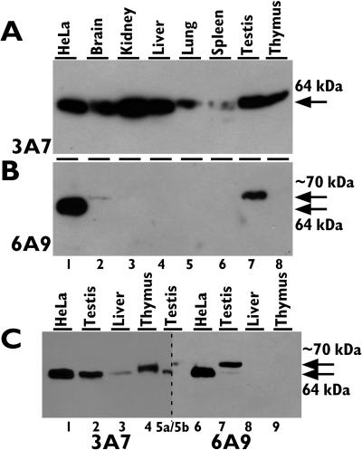
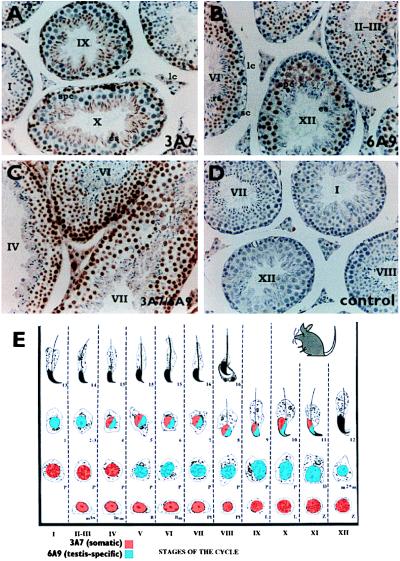
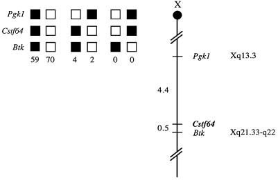
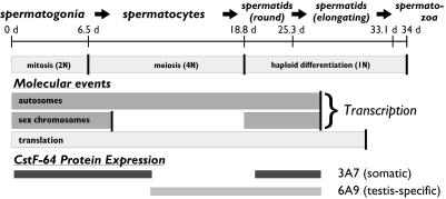
Polyadenylation in male germ cells differs from that in somatic cells. Many germ cell mRNAs do not contain the canonical AAUAAA in their 3' ends but are efficiently polyadenylated. To determine whether the 64,000 Mr protein of the cleavage stimulation factor (CstF-64) is altered in male germ cells, we examined its expression in mouse testis. In addition to the 64,000 Mr form, we found a related approximately 70,000 Mr protein that is abundant in testis, at low levels in brain, and undetectable in all other tissues examined. Expression of the approximately 70,000 Mr CstF-64 was limited to meiotic spermatocytes and postmeiotic spermatids in testis. In contrast, the 64,000 Mr form was absent from spermatocytes, suggesting that the testis-specific CstF-64 might control expression of meiosis-specific genes. To determine why the 64,000 Mr CstF-64 is not expressed in spermatocytes, we mapped its chromosomal location to the X chromosome in both mouse and human. CstF-64 may, therefore, be absent in spermatocytes because the X chromosome is inactivated during male meiosis. By extension, the testis-specific CstF-64 may be expressed from an autosomal homolog of the X chromosomal gene.
Figures

Similar articles
-
The gene for a variant form of the polyadenylation protein CstF-64 is on chromosome 19 and is expressed in pachytene spermatocytes in mice.J Biol Chem. 2001 Mar 16;276(11):8044-50. doi: 10.1074/jbc.M009091200. Epub 2000 Dec 11. J Biol Chem. 2001. PMID: 11113135
-
Elevated levels of the polyadenylation factor CstF 64 enhance formation of the 1kB Testis brain RNA-binding protein (TB-RBP) mRNA in male germ cells.Mol Reprod Dev. 2001 Apr;58(4):460-9. doi: 10.1002/1098-2795(20010401)58:4<460::AID-MRD15>3.0.CO;2-F. Mol Reprod Dev. 2001. PMID: 11241784
-
Developmental distribution of the polyadenylation protein CstF-64 and the variant tauCstF-64 in mouse and rat testis.Biol Reprod. 2004 Apr;70(4):1080-7. doi: 10.1095/biolreprod.103.022947. Epub 2003 Dec 17. Biol Reprod. 2004. PMID: 14681198
-
Overexpression of the CstF-64 and CPSF-160 polyadenylation protein messenger RNAs in mouse male germ cells.Biol Reprod. 2001 Jun;64(6):1722-9. doi: 10.1095/biolreprod64.6.1722. Biol Reprod. 2001. PMID: 11369601
-
The gene CSTF2T, encoding the human variant CstF-64 polyadenylation protein tauCstF-64, lacks introns and may be associated with male sterility.Genomics. 2002 Nov;80(5):509-14. Genomics. 2002. PMID: 12408968
Cited by
-
The end of the message: multiple protein-RNA interactions define the mRNA polyadenylation site.Genes Dev. 2015 May 1;29(9):889-97. doi: 10.1101/gad.261974.115. Genes Dev. 2015. PMID: 25934501 Free PMC article. Review.
-
Calmodulin interacts with and regulates the RNA-binding activity of an Arabidopsis polyadenylation factor subunit.Plant Physiol. 2006 Apr;140(4):1507-21. doi: 10.1104/pp.105.070672. Epub 2006 Feb 24. Plant Physiol. 2006. PMID: 16500995 Free PMC article.
-
CstF-64 supports pluripotency and regulates cell cycle progression in embryonic stem cells through histone 3' end processing.Nucleic Acids Res. 2014 Jul;42(13):8330-42. doi: 10.1093/nar/gku551. Epub 2014 Jun 23. Nucleic Acids Res. 2014. PMID: 24957598 Free PMC article.
-
Molecular architecture of the human pre-mRNA 3' processing complex.Mol Cell. 2009 Feb 13;33(3):365-76. doi: 10.1016/j.molcel.2008.12.028. Mol Cell. 2009. PMID: 19217410 Free PMC article.
-
Transcriptome-wide analyses of CstF64-RNA interactions in global regulation of mRNA alternative polyadenylation.Proc Natl Acad Sci U S A. 2012 Nov 13;109(46):18773-8. doi: 10.1073/pnas.1211101109. Epub 2012 Oct 29. Proc Natl Acad Sci U S A. 2012. PMID: 23112178 Free PMC article.
References
Publication types
MeSH terms
Substances
Grants and funding
LinkOut - more resources
Full Text Sources
Other Literature Sources
Molecular Biology Databases
