. 1999 Jun 8;96(12):6791-6.
doi: 10.1073/pnas.96.12.6791.

A quantitative analysis of signal transduction from activin receptor to nucleus and its relevance to morphogen gradient interpretation

Affiliations

A quantitative analysis of signal transduction from activin receptor to nucleus and its relevance to morphogen gradient interpretation

K Shimizu et al. Proc Natl Acad Sci U S A. .

Abstract

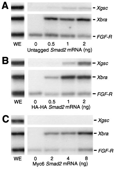
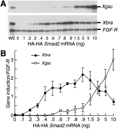
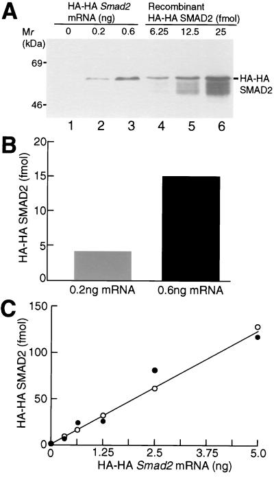
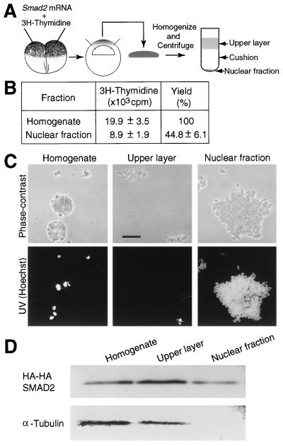
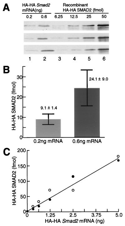
Previous work has shown that Xenopus blastula cells sense activin concentration by assessing the absolute number of occupied receptors per cell (100 and 300 molecules of bound activin activate Xbra and Xgsc transcription, respectively; a difference of only 3-fold). We now ask how quantitative differences in the absolute number of occupied receptors lead to the qualitatively distinct gene responses in the nucleus through SMAD2, a transducer of concentration-dependent gene responses to activin. We show that the injection of 0.2 or 0.6 ng of Smad2 mRNA activates Xbra or Xgsc transcription, respectively, involving, again, only a 3-fold difference. Furthermore, Xbra transcription is down-regulated by overexpression of SMAD2 as it is after activin signaling. We have developed a method to isolate nuclei from animal cap cells and subsequently have quantified the amount of nuclear SMAD2 protein. We find that the injection of 0.2 or 0.6 ng of Smad2 mRNA into an egg leads to only a 3-fold difference in the amount of SMAD2 protein in the nuclei of the blastula cells that express Xbra or Xgsc. We conclude that a 3-fold difference in the absolute number of occupied activin receptors can be maintained only as a 3-fold difference in the level of nuclear SMAD2 protein. Therefore, in this example of morphogen action, there appears to be no amplification of a key cytoplasmic transduction response, and a small but developmentally important change in extracellular signal concentration is relayed directly to the nucleus.


Figures

Figure 1
Figure 1
Xbra and Xgsc transcription is activated by HA-HA SMAD2 as effectively as by untagged SMAD2. Each embryo was injected with the indicated dose of untagged, HA-HA, or Myc6 Smad2 mRNA. Animal caps at stage 10.5 were analyzed by RNase protection. WE, whole embryo. The results shown are representative of three independent experiments.
Figure 2
Figure 2
Activation of Xbra and Xgsc transcription by HA-HA SMAD2 in the same dose-responsive way as activin. (A) RNase protection analysis of Xbra and Xgsc expression. Each embryo was injected with the indicated dose of HA-HA Smad2 mRNA. Animal caps at stage 10.5 were analyzed by RNase protection. WE, whole embryo. The result shown is representative of three independent experiments. (B) Quantitation of gel analyses. The levels of Xbra and Xgsc transcription relative to fibroblast growth factor receptor loading control were quantified by a Bio-imaging analyzer, BAS-2500. The values are means ± SEs of three independent experiments.
Figure 3
Figure 3
Quantitation of the amount of HA-HA SMAD2 protein in animal cap cells. (A) Western blotting of HA-HA SMAD2. Each embryo was injected with the indicated dose of HA-HA Smad2 mRNA. Animal caps were homogenized at stage 10.5. Twelve micrograms of total protein from mRNA-injected embryos (lanes 1–3) or the indicated amount of recombinant HA-HA SMAD2 protein (lanes 4–6) was subjected to SDS/PAGE, followed by Western blotting with the anti-HA antibody. The result shown is representative of three independent experiments. (B) Quantitation of the amount of HA-HA SMAD2 protein. The amounts of HA-HA SMAD2 protein in lanes 2 and 3 of A were quantified by using a standard curve based on recombinant HA-HA SMAD2 protein. (C) Quantitation of the amount of HA-HA SMAD2 protein synthesized by embryos injected with various doses of HA-HA Smad2 mRNA. Each embryo was injected with the indicated dose of HA-HA Smad2 mRNA. The amount of HA-HA SMAD2 protein per 12 μg of total protein was quantified as described in B. The results shown are drawn from two independent experiments. In a third experiment, all points were somewhat higher, but fell on a straight line.
Figure 4
Figure 4
Preparation of a nuclear fraction. (A) Strategy to isolate nuclei. (B) The yield of nuclei. Fifty embryos were injected with [3H]thymidine (8,500 cpm/embryo). The acid-insoluble radioactivity of the homogenate and the nuclear fraction was scintillation counted. The values are means ± SEs of five independent experiments. (C) Photographs of subfractions. The homogenate, the upper layer, and the nuclear fraction were observed and photographed under phase-contrast or UV illumination. (Bar = 0.1 mm.) (D) Western blotting of subfractions. Each embryo was injected with HA-HA Smad2 mRNA (1 ng) and with [3H]thymidine (8,500 cpm). The homogenate (10 μg of total protein), the upper layer (10 μg of total protein), equivalent to about one-third of an animal cap, and the nuclear fraction containing acid-insoluble 1,000 cpm of [3H]thymidine, equivalent to about two animal caps, were subjected to SDS/PAGE, followed by Western blotting with the anti-HA or anti-α-tubulin antibodies. The results shown in C and D are representative of three independent experiments.
Figure 5
Figure 5
Quantitation of the amount of HA-HA SMAD2 protein in the nuclei. (A) Western blotting of HA-HA SMAD2 protein. Each embryo was injected with 0.2 or 0.6 ng of HA-HA Smad2 mRNA and with 8,500 cpm of [3H]thymidine. Protein, from the nuclear fractions of mRNA-injected embryos, containing 6,700 cpm of acid-insoluble [3H]thymidine (lanes 1 and 2) or the indicated amount of recombinant HA-HA SMAD2 protein (lanes 3–6) was subjected to SDS/PAGE, followed by Western blotting with the anti-HA antibody. The result shown are from three independent experiments. (B) Quantitation of the amount of HA-HA SMAD2 protein. The amounts of HA-HA SMAD2 in lanes 1 and 2 of A were quantified by using a standard curve based on recombinant HA-HA SMAD2 protein. The values are means ± SEs of three independent experiments. (C) Quantitation of the amount of HA-HA SMAD2 protein synthesized by embryos injected with various doses of HA-HA Smad2 mRNA. Each embryo was injected with the indicated dose of Smad2 mRNA. The amount of HA-HA SMAD2 protein in the nuclear fraction containing 6,700 cpm of acid-insoluble [3H]thymidine was quantified as described in Fig. 5B. The results shown are drawn from two independent experiments. In a third experiment, all points were higher, but fell on a straight line.

References

    1. Wolpert L. J Theor Biol. 1969;25:1–47. - PubMed
    1. Gurdon J B, Dyson S, St. Johnston D. Cell. 1998;95:159–162. - PubMed
    1. Lawrence P A, Struhl G. Cell. 1996;85:951–961. - PubMed
    1. Neumann C, Cohen S. BioEssays. 1997;19:721–729. - PubMed
    1. Green J B A, Smith J C. Nature (London) 1990;347:391–394. - PubMed

Publication types

LinkOut - more resources