

CCR5(+) and CXCR3(+) T cells are increased in multiple sclerosis and their ligands MIP-1alpha and IP-10 are expressed in demyelinating brain lesions
- PMID: 10359806
- PMCID: PMC22009
- DOI: 10.1073/pnas.96.12.6873
CCR5(+) and CXCR3(+) T cells are increased in multiple sclerosis and their ligands MIP-1alpha and IP-10 are expressed in demyelinating brain lesions
Abstract
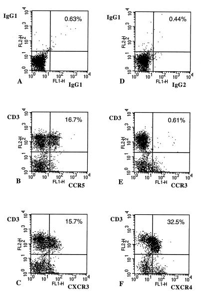
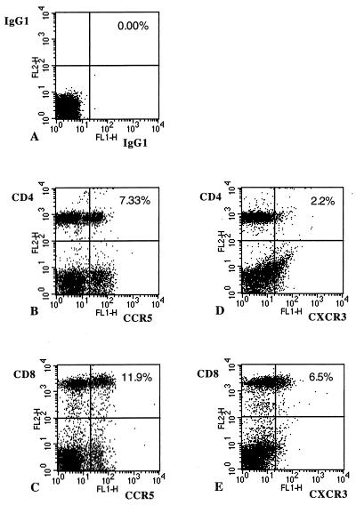
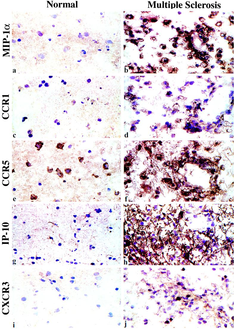
Multiple sclerosis (MS) is a T cell-dependent chronic inflammatory disease of the central nervous system. The role of chemokines in MS and its different stages is uncertain. Recent data suggest a bias in expression of chemokine receptors by Th1 vs. Th2 cells; human Th1 clones express CXCR3 and CCR5 and Th2 clones express CCR3 and CCR4. Chemokine receptors expressed by Th1 cells may be important in MS, as increased interferon-gamma (IFN-gamma) precedes clinical attacks, and IFN-gamma injection induces disease exacerbations. We found CXCR3(+) T cells increased in blood of relapsing-remitting MS, and both CCR5(+) and CXCR3(+) T cells increased in progressive MS compared with controls. Furthermore, peripheral blood CCR5(+) T cells secreted high levels of IFN-gamma. In the brain, the CCR5 ligand, MIP-1alpha, was strongly associated with microglia/macrophages, and the CXCR3 ligand, IP-10, was expressed by astrocytes in MS lesions but not unaffected white matter of control or MS subjects. Areas of plaque formation were infiltrated by CCR5-expressing and, to a lesser extent, CXCR3-expressing cells; Interleukin (IL)-18 and IFN-gamma were expressed in demyelinating lesions. No leukocyte expression of CCR3, CCR4, or six other chemokines, or anti-inflammatory cytokines IL-5, IL-10, IL-13, and transforming growth factor-beta was observed. Thus, chemokine receptor expression may be used for immunologic staging of MS and potentially for other chronic autoimmune/inflammatory processes such as rheumatoid arthritis, autoimmune diabetes, or chronic transplant rejection. Furthermore, these results provide a rationale for the use of agents that block CCR5 and/or CXCR3 as a therapeutic approach in the treatment of MS.
Figures
References
-
- Luster A D. New Engl J Med. 1998;338:436–445. - PubMed
-
- Sallusto F, Mackay C R, Lanzavecchia A. Science. 1997;277:2005–2007. - PubMed
-
- Zingoni A, Soto H, Hedrick J A, Stoppacciaro A, Storlazzi C T, Sinigaglia F, D’Ambrosio D, Ogarra A, Robinson D, Rocchi M, et al. J Immunol. 1998;161:547–551. - PubMed
Publication types
MeSH terms
Substances
Grants and funding
LinkOut - more resources
Full Text Sources
Other Literature Sources
Medical
Miscellaneous
