. 1999 Jun 8;96(12):6879-84.
doi: 10.1073/pnas.96.12.6879.

Broad tumor-associated expression and recognition by tumor-derived gamma delta T cells of MICA and MICB

Affiliations

Broad tumor-associated expression and recognition by tumor-derived gamma delta T cells of MICA and MICB

V Groh et al. Proc Natl Acad Sci U S A. .

Abstract

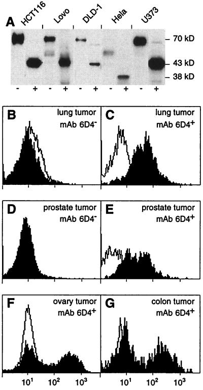
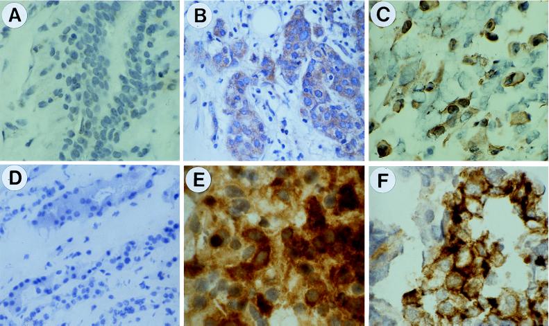
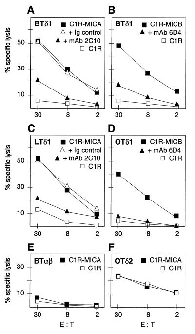
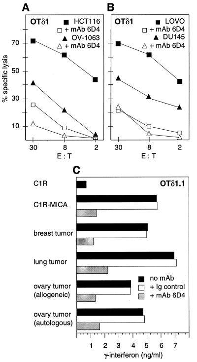
Human MHC class I-related molecules, MICA and MICB, are stress-induced antigens that are recognized by a subset of gamma delta T cells expressing the variable region Vdelta1. This functional association has been found to be limited to intestinal epithelium, where these T cells are prevalent and where MICA and, presumably, MICB are mainly expressed. However, increased frequencies of Vdelta1 gamma delta T cells have been observed in various epithelial tumors; moreover, MICA/B are expressed on diverse cultured epithelial tumor cells. With freshly isolated tumor specimens, expression of MICA/B was documented in many, but not all, carcinomas of the lung, breast, kidney, ovary, prostate, and colon. In tumors that were positive for MICA/B, the frequencies of Vdelta1 gamma delta T cells were significantly higher than in those that were negative. Vdelta1 gamma delta T cell lines and clones derived from different tumors recognized MICA/B on autologous and heterologous tumor cells. In accord with previous evidence, no constraints were observed in these interactions, such as those imposed by specific peptide ligands. Thus, MICA/B are tumor-associated antigens that can be recognized, in an apparently unconditional manner, by a subset of tumor-infiltrating gamma delta T cells. These results raise the possibility that an induced expression of MICA/B, by conditions that may be related to tumor homeostasis and growth, could play a role in immune responses against tumors.


Figures

Figure 1
Figure 1
Surface expression of MICA on epithelial tumor cell lines and of MICA/B on freshly isolated tumor cells. (A) Immunoprecipitation of MICA with mAb 2C10 from lysates of surface-biotinylated HCT116, Lovo, DLD-1 (colon carcinomas), Hela (cervical carcinoma), and U373 (astrocytoma) tumor cell lines. Immunocomplexes were left untreated (−) or were treated (+) with N-glycanase before SDS/PAGE. Labeled proteins on immunoblots were visualized with avidin-horseradish peroxidase. MICA is heavily glycosylated and has a polypeptide backbone of 43 kDa (11, 13). These results were in accord with previous data obtained with the anti-MICA mAb 56 by using transfectants and epithelial cell lines (13). Hela cells are homozygous for a truncated variant of MICA of 38 kDa that lacks the cytoplasmic tail. This variant corresponds to allele 008, as determined by reverse transcription–PCR and direct cDNA sequencing (unpublished data) (24). (B–G) Tumor cell suspensions prepared from lung (B and C), prostate (D and E), ovary (F), and colon carcinomas (G) were stained with mAb 6D4 (solid profiles) or with isotype-matched Ig (open profiles) and examined by flow cytometry. B and D show examples of tumor cell preparations that were negative for binding of mAb 6D4 whereas C and EG show positive stainings. These data are representative of the range of staining intensities seen with all of the tumor samples tested. Similar results were obtained with mAb 2C10.
Figure 2
Figure 2
Immunohistochemical detection of MICA/B in epithelial tumors by staining with mAb 6D4. (A) Normal breast tissue. (B) Breast carcinoma. (C) Lung carcinoma. (D) Normal kidney. (E) Renal cell carcinoma. (F) Ovary carcinoma. Sections in A and B and CF were from paraffin-embedded and frozen specimens, respectively. Nuclei were counterstained with hematoxylin (see Materials and Methods). These images were representative of stainings of numerous control tissue and tumor specimens. See text for further explanation.
Figure 3
Figure 3
Specific recognition of MICA/B on C1R transfectants by the Vδ1 γδ T cell lines BTδ1, LTδ1, and OTδ1 derived from carcinomas of breast, lung, and ovary, respectively (AD). The data from chromium release assays shown were representative of numerous experiments with these T cell lines and others derived from prostate and colon carcinomas, which all were tested against the C1R-MICA and C1R-MICB targets. Cytotoxicity was inhibited in the presence of mAb 2C10 (anti-MICA) or mAb 6D4 (anti-MICA and -MICB) but not with control Ig. (E) No responses were seen with the tumor-derived CD8+ αβ T cell lines; as one example, data obtained with the breast tumor-derived BTαβ line are shown. (F) The ovary tumor-derived Vδ2 γδ T cell line (OTδ2) showed no increased response against C1R-MICA targets. E:T, effector-to-target cell ratio. See text for further explanation.
Figure 4
Figure 4
Vδ1 γδ T cells recognized MICA/B on epithelial tumor cell lines and on heterologous and autologous tumor cells. (A and B) Cytotoxicity of the ovary tumor-derived OTδ1 T cell line against HCT116 and Lovo (colon carcinoma cell lines), OV-1063 (ovary carcinoma cell line), and DU145 (prostate carcinoma line) was inhibited by mAb 6D4. Addition of isotype-matched control Ig had no effect (data not shown). The target cell lines expressed MICA/B by staining with mAb 6D4 (ref. ; data not shown). Similar results were obtained with other tumor-derived Vδ1 γδ T cell lines and additional tumor cell line targets (see text). E:T, effector-to-target cell ratio. (C) MICA/B-induced release of IFN-γ by the OTδ1.1 T cell clone was inhibited in the presence of mAb 6D4 but not of isotype-matched control Ig. Release of IFN-γ (ng/ml) after 48 h of T cell stimulation was quantitated by ELISA (see Materials and Methods). Stimulator cells were C1R-MICA transfectants or tumor cell preparations from breast, lung, or heterologous or autologous ovary carcinomas. Similar results were obtained with another Vδ1 γδ T cell clone derived from a different ovary tumor.

Similar articles

Cited by

References

    1. Van den Eynde B J, van der Bruggen P. Curr Opin Immunol. 1997;9:684–693. - PubMed
    1. Pamer E, Cresswell P. Annu Rev Immunol. 1998;16:323–358. - PubMed
    1. Rammensee H-G, Falk K, Ràtzschke O. Annu Rev Immunol. 1993;11:213–244. - PubMed
    1. Ferrone S, Marincola F M. Immunol Today. 1995;16:487–494. - PubMed
    1. Allison J P, Raulet D H. Semin Immunol. 1990;2:59–65. - PubMed

Publication types

MeSH terms

Substances