. 1999 Jun 8;96(12):7023-8.
doi: 10.1073/pnas.96.12.7023.

Calexcitin transformation of GABAergic synapses: from excitation filter to amplifier

Affiliations

Calexcitin transformation of GABAergic synapses: from excitation filter to amplifier

M K Sun et al. Proc Natl Acad Sci U S A. .

Abstract

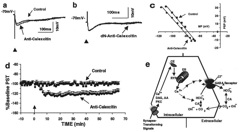
Encoding an experience into a lasting memory is thought to involve an altered operation of relevant synapses and a variety of other subcellular processes, including changed activity of specific proteins. Here, we report direct evidence that co-applying (associating) membrane depolarization of rat hippocampal CA1 pyramidal cells with intracellular microinjections of calexcitin (CE), a memory-related signaling protein, induces a long-term transformation of inhibitory postsynaptic potentials from basket interneurons (BAS) into excitatory postsynaptic potentials. This synaptic transformation changes the function of the synaptic inputs from excitation filter to amplifier, is accompanied by a shift of the reversal potential of BAS-CA1 postsynaptic potentials, and is blocked by inhibiting carbonic anhydrase or antagonizing ryanodine receptors. Effects in the opposite direction are produced when anti-CE antibody is introduced into the cells, whereas heat-inactivated CE and antibodies are ineffective. These data suggest that CE is actively involved in shaping BAS-CA1 synaptic plasticity and controlling information processing through the hippocampal networks.


Figures

Figure 1
Figure 1
CE transforms BAS–CA1 synapses. Bicuculline (BIC, 1 μM, 30 min) eliminates (a), whereas KYN (500 μM, 20 min) does not alter (b), the evoked IPSPs. The relationship between the evoked BAS–CA1 PSP at different membrane potentials (MPs) (c and d) in a CA1 pyramidal cell can be described with a straight line (e), determined by the least sum squares criterion, and is not altered by KYN (c and d). CE reduces BAS–CA1 IPSP (f; two overlapping traces) and shifts the PSP–MP curve to the right (h). Heat-inactivated CE (dN-Calexcitin) is ineffective (g). Microinjections of CE conjugated with the green fluorescent Alexa488 (Molecular Probes) results in strong labeling of the cell body and portion of the dendrites in focus (i; Left, active form, and Right, heat-inactivated; after fixation with 10% paraformaldehyde/saline overnight and cutting to 40 μm thick, shown ×400), indicating the efficacy of the CE microinjection. In j, time courses of the response to CE or heat-inactivated CE injection (Control), each point represents the mean IPSP magnitudes + SEM normalized to the average of the pre-CE IPSPs. PST, postsynaptic transformation. The vertical arrow indicates the time of injection. Associating CE injection with postsynaptic depolarization (0.4–0.6 nA during the off period with the current intensities adjusted to elicit 4–8 spikes per s) transforms BAS–CA1 inhibitory PSP into an excitatory one (k) and produces a further shift of the PSP–MP curve to the right (m). Associating heat-inactivated CE with postsynaptic depolarization (0.4–0.8 nA at the off period with current intensities adjusted to evoke 4–8 spikes per s) does not alter BAS–CA1 IPSP (L). Average responses of BAS–CA1 PSP after associating either CE (CE+Ca2+) or heat-inactivated CE (Control) are shown in n. The transformed synaptic response is eliminated by 1 μM bicuculline (o; 30 min).
Figure 2
Figure 2
Anti-CE antibody enhances BAS–CA1 IPSPs and mechanisms of CE-induced transformation of GABAergic synapses. Anti-CE antibody injection into a recorded CA1 pyramidal cell enhances BAS–CA1 IPSP (a, as compared with unmarked IPSP before injection) and elicits a shift of the PSP–MP curve to the left (c). Injection of heat-inactivated antibody is ineffective (b; two traces overlapping). Average responses of BAS–CA1 PSP after injection of either anti-CE antibody (Anti-CE) or its heat-inactivated form (Control) are shown in d. Schematic drawing (e) shows mechanisms of CE-mediated transformation of GABAergic synapses. Synapse-transforming signals (such as associative activation of cholinergic and GABAergic inputs) turn on a CE/CE-like protein signal cascade. CE binds to the RyR and causes Ca2+ release. The Ca2+/CE transforms the GABAergic synapses by shifting the GABAA reversal potential from Cl reversal potential toward HCO3 reversal potential, through altering anion selectivity of the Cl channels, activity of CA, and/or formation of HCO3. Multiple arrows indicate possible involvement of unidentified mediators. AA, arachidonic acid; DAG, diacylglycerol; ER, endoplasmic reticulum; PKC, protein kinase C.
Figure 3
Figure 3
ACET and non-bicarbonate buffer eliminate CE-induced transformation, and the transformation converts excitatory input filter into amplifier. ACET (1 μM) eliminates CE-induced synaptic transformation (a). The effect of CE on BAS–CA1 IPSPs is not observed in Hepes buffer (b). In the presence of extracellular benzolamide (10 μM), CE depolarization induces the synaptic transformation (c), which is not induced when BAPTA is co-applied (d; the charges carried by BAPTA are compensated by reducing the amount of acetate). Single-pulse stimulation (e) of BAS–CA1 evokes an IPSP and of SCH at above-threshold intensities, action potentials (truncated; two traces: one stimulated at delay of 10 ms and the other 30 ms, marked with arrows). The excitatory SCH (at the same above-threshold stimulation) input is filtered out by a costimulation of BAS–CA1 (f; two overlapping traces). (g) Single-pulse stimulation of BAS–CA1 evokes an IPSP, and stimulation of SCH at below-threshold intensities evokes an EPSP. The excitatory SCH (at the same below-threshold stimulation) input is below threshold as evoked by costimulation (single pulse) of BAS–CA1 and SCH inputs (h) before CE application. CE (30 min after the application) transforms BAS–CA1 IPSP and does not change much of the SCH–CA1 EPSP, evoked by single-pulse stimulation of BAS or SCH, respectively (i). The excitatory SCH (at the same below-threshold stimulation) input is amplified by the co-BAS stimulation after the CE-induced synaptic transformation and induces action potentials (truncated; j: two overlapping traces). (k) Schematic diagram of transformed GABAergic synapse functioning as either excitatory filter (surround) or amplifier (center). Active BAS GABAergic inputs effectively filter excitatory signals so that only very strong excitatory inputs might evoke action potentials. The GABAergic synaptic transformation results in amplifying excitatory signals so that weaker inputs can pass through the neural circuits (through the cell in the middle). BAS, basket GABAergic interneurons (in black); Pyr, CA1 pyramidal cells.

References

    1. Kornhauser J M, Greenberg M E. Neuron. 1997;18:839–842. - PubMed
    1. Alkon D L, Nelson T J, Zhao W Q, Cavallaro S. Trends Neurosci. 1998;21:529–537. - PubMed
    1. Nelson T J, Collin C, Alkon D L. Science. 1990;247:1479–1483. - PubMed
    1. Sun M-K. Pharmacol Rev. 1996;48:465–494. - PubMed
    1. McMahon L L, Kauer J A. Neuron. 1997;18:295–305. - PubMed

Substances

LinkOut - more resources