. 1999 Jun 8;96(12):7071-6.
doi: 10.1073/pnas.96.12.7071.

Isolation and characterization of a dual-substrate phosphodiesterase gene family: PDE10A

Affiliations

Isolation and characterization of a dual-substrate phosphodiesterase gene family: PDE10A

S H Soderling et al. Proc Natl Acad Sci U S A. .

Abstract

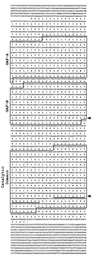
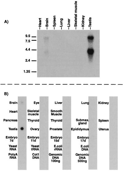
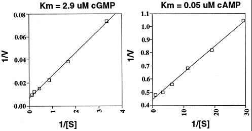
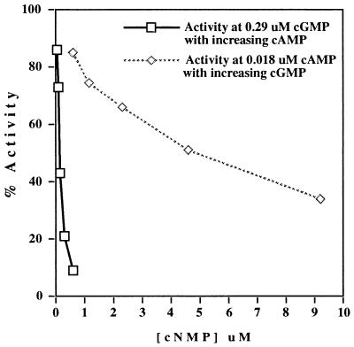
We report here the cloning, expression, and characterization of a dual-substrate, cAMP and cGMP, cyclic nucleotide phosphodiesterase (PDE) from mouse. This PDE contains the consensus sequence for a PDE catalytic domain, but shares <50% sequence identity with the catalytic domains of all other known PDEs and, therefore, represents a new PDE gene family, designated PDE10A. The cDNA for PDE10A is 3, 370 nt in length. It includes a full ORF, contains three in-frame stop codons upstream of the first methionine, and is predicted to encode a 779-aa enzyme. At the N terminus PDE10A has two GAF domains homologous to many signaling molecules, including PDE2, PDE5, and PDE6, which likely constitute a low-affinity binding site for cGMP. PDE10A hydrolyzes cAMP with a Km of 0.05 microM and cGMP with a Km of 3 microM. Although PDE10A has a lower Km for cAMP, the Vmax ratio (cGMP/cAMP) is 4.7. RNA distribution studies indicate that PDE10A is expressed at highest levels in testis and brain.


Figures

Figure 1
Figure 1
Full-length sequence of MMPDE10A. cDNA sequence of PDE10A is shown. Bold codons above the predicted start methionine are in-frame stop codons indicating the cDNA contains the entire ORF. Boxed amino acid regions indicate domains identified by sequence similarity to known domains in other proteins. Bold, italic amino acids in GAFA and GAFB domains correspond to residues thought to be important for the support of cGMP binding in PDE5. Top arrow points to the consensus motif [N(K/R)XnFX3DE] found in full in GAFB. Lower arrow points to a predicted bipartite nuclear localization signal found within the C terminus of PDE10A. Asterisk indicates the final stop codon.
Figure 2
Figure 2
Northern blot and RNA dot blot analyses of PDE10A expression. (A) Northern blot analysis of poly(A) RNA from mouse tissues. Two bands, 9 kb and 4 kb, are detected in both testis and brain that hybridize to PDE10A probes. (B) RNA dot blot analysis of 22 mouse tissues. Consistent with Northern data, mouse testis and brain appear to express PDE10A, with highest levels of expression in testis.
Figure 3
Figure 3
PDE10A kinetics. Shown are Lineweaver–Burk plots for both cAMP and cGMP. The displayed Km is the calculated average number.
Figure 4
Figure 4
cGMP-binding assays for PDE2 and PDE10A. Binding is detected for PDE2 at 9.2 μM, whereas much less binding is detected for PDE10A. Results are average picomoles bound, with range shown for PDE2 (n = 2) and SD shown for PDE10 (n = 6).
Figure 5
Figure 5
Effect of cGMP on cAMP hydrolysis and effect of cAMP on cGMP hydrolysis. [3H]cAMP hydrolysis was measured in the presence of 0.018 μM cAMP and a range of cGMP concentrations from 0.6 to 9.2 μM. The reverse also was done with [3H]cGMP (and 0.29 μM cGMP) and 0.052–0.6 μM cAMP. cAMP may function as a potent inhibitor of cGMP hydrolysis.
Figure 6
Figure 6
Multiple sequence alignment of the C-terminal PDE10A GAF domains to GAF domains of other proteins. Numbers in parentheses at the end of the sequences indicate the amino acid numbers shown for each protein. Numbers in parentheses within sequence alignment indicate the number of amino acids omitted from the alignment for clarity. Arrows mark the motif described as supporting cGMP binding in PDE5. Shading reflects at least 50% conservation. BTPDE2, Bos taurus PDE2 (accession no. M73512); HSPDE5, Homo sapiens PDE5 (AF043731); CFPDE6A, Canis familiaris PDE6 alpha (Y13282); EcfhlA, E. coli FhlA (X52227); CEPDE, C. elegans gene R0807.6 “similar to phosphodiesterase” (Z12017); YEykl0, Saccharomyces cerevisiae ORF YKL069W (Z28069.1); AtphyE, Arabidopsis thaliana phytochrome E (X76610); AFHisK, Archaeoglobus fulgidus gene AF1483 “putative histidine kinase” (AE001000).

References

    1. Beavo J A. Physiol Rev. 1995;75:725–748. - PubMed
    1. Zhao A Z, Zhao H, Teague J, Fujimoto W, Beavo J A. Proc Natl Acad Sci USA. 1997;94:3223–3228. - PMC - PubMed
    1. Zhao A Z, Bornfeldt K E, Beavo J A. J Clin Invest. 1998;102:869–873. - PMC - PubMed
    1. Li L, Yee C, Beavo J A. Science. 1999;283:848–851. - PubMed
    1. Ballard S A, Gingell C J, Tang K, Turner L A, Price M E, Naylor A M. J Urol. 1998;159:2164–2171. - PubMed

Publication types

MeSH terms

Associated data