. 1999 Jun;116(3):453-61.
doi: 10.1046/j.1365-2249.1999.00904.x.

Porins and lipopolysaccharide (LPS) from Salmonella typhimurium induce leucocyte transmigration through human endothelial cells in vitro

Affiliations

Porins and lipopolysaccharide (LPS) from Salmonella typhimurium induce leucocyte transmigration through human endothelial cells in vitro

M Galdiero et al. Clin Exp Immunol. 1999 Jun.

Expression of concern in

Abstract

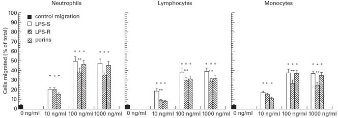
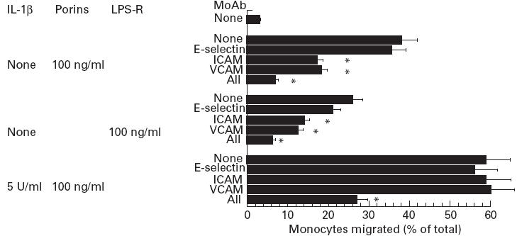
Bacteria or bacterial products may constitute important inducers of surface molecule expression on endothelial cells and leucocytes. This study was undertaken to determine the effects of the Salmonella typhimurium porins, LPS-S and LPS-R on the transendothelial migration of leucocytes through human umbilical vein endothelial cells (HUVEC). Treatment of the HUVEC with either porins or LPS-S or LPS-R increased the transmigration of different leucocyte populations, in particular that of neutrophils. The maximal increase occurred using LPS-S treatment, whereas porin stimulation fell between LPS-S and LPS-R. The transmigration increase was dose-dependent and reached its maximum at about 100-1000 ng/ml of stimulus. Optimal endothelial activation occurred after 2-4 h and 4-6 h using LPS and porin, respectively. Stimulation of leucocytes with either porins or LPS slightly increased their transmigration through non-activated endothelial cells. Transmigration increased remarkably during the simultaneous stimulation of endothelial cells by IL-1ss together with either porins or LPS. To assess participation of E-selectin, intercellular adhesion molecule-1 (ICAM-1), vascular cell adhesion molecule-1 (VCAM-1) and leucocyte adhesion complex (CD11/18) in porin- or LPS-mediated leucocyte migration, blocking MoAbs were used. Each blocking MoAb partially and selectively decreased leucocyte transmigration. The obtained results contribute to clarify some aspects of the inflammatory process at sites of infection.


Figures

Fig. 1
Fig. 1
SDS–PAGE analysis of porins and LPS preparations from Salmonella typhimurium. Lane A, molecular weight standards (phosphorylase b, 94 000; albumin, 67 000; ovalbumin, 43 000; carbonic anydrase, 30 000; trypsin inhibitor, 20 100; α-lactalbumina, 14 000). Lane B, SH5014 porins. Lane C, SH5014 LPS-R. Lane D, 74 NCTC LPS.
Fig. 2
Fig. 2
Porin-, LPS-S-, LPS-R-stimulated migration of leucocytes across human umbilical vein endothelial cells (HUVEC) cultured on human fibronectin-coated culture insert (6.4 mm diameter) in a dose-dependent manner. Monolayers of HUVEC were incubated with either medium (control) or LPS for 4 h or porins for 6 h and washed; leucocytes were then added and allowed to migrate for 2 h. Data represent the mean ± s.e.m. of three independent experiments. *P < 0.001 versus control migration; **P < 0.05 versus LPS-S-stimulated leucocytes.
Fig. 3
Fig. 3
Time course of porin-, LPS-S- and LPS-R-induced leucocyte transendothelial migration. Human umbilical vein endothelial cells (HUVEC) were incubated with either medium (control, ▴) or 100 ng/ml of stimulus for 2 h and at the indicated time periods leucocytes were added and allowed to migrate for 2 h. □, LPS-S; •, porins; ○, LPS-R. Data shown are mean ± s.e.m. of three independent experiments. (P < 0.001 comparison with control migration).
Fig. 4
Fig. 4
Transendothelial migration of stimulated leucocytes across non- stimulated human umbilical vein endothelial cells (HUVEC). Leucocytes were treated separately with stimuli (100 ng/1.5 × 106 cells/ml) for 2 h, were then washed and added to non-stimulated monolayer. The data represent mean ± s.e.m. of three independent experiments. *P < 0.001 versus control migration; **P < 0.05 versus LPS-S-stimulated leucocytes.
Fig. 5
Fig. 5
Migration of leucocytes across endothelial monolayer activated by IL-1ß for 4 h and then stimulated by porins, LPS-S and LPS-R for 2 h. The data represent mean ± s.e.m. of three independent experiments.
Fig. 6
Fig. 6
Migration inhibition of neutrophils across human umbilical vein endothelial cell (HUVEC) monolayer by pretreatment with MoAb for 45 min. Monolayers of HUVEC were incubated with either IL-1ß for 4 h or porins for 6 h or LPS-R for 4 h. In each separate experiment the control MoAbs (W27, BL-1a/DQ) of the same isotype of MoAbs used had no significant effect on porin- or LPS-R-stimulated migration. Results are representative of three independent experiments; bars, mean ± s.e.m.; *P < 0.001.
Fig. 7
Fig. 7
Migration inhibition of monocytes across human umbilical vein endothelial cell (HUVEC) monolayer by pretreatment with MoAbs for 45 min. Monolayers of HUVEC were incubated with either IL-1ß for 4 h or porins for 6 h or LPS-R for 4 h. In each separate experiment the control MoAbs (W27, BL-1a/DQ) of the same isotype of MoAbs used had no significant effect on porin- or LPS-R-stimulated migration. Results are representative of three independent experiments; bars, mean ± s.e.m.; *P < 0.001.
Fig. 8
Fig. 8
Migration inhibition of lymphocytes across human umbilical vein endothelial cell (HUVEC) monolayer by pretreatment with MoAbs for 45 min. Monolayers of HUVEC were incubated with either IL-1ß for 4 h or porins for 6 h or LPS-R for 4 h. In each separate experiment the control MoAbs (W27, BL-1a/DQ) of the same isotype of MoAb used had no significant effect on porin- or LPS-R-stimulated migration. Results are representative of three independent experiments; bars, mean ± s.e.m.; *P < 0.001.
Fig. 9
Fig. 9
Migration inhibition across human umbilical vein endothelial cell (HUVEC) monolayer activated by IL-1ß (5 U/ml) of leucocytes pretreated with MoAbs for 45 min. Monolayers of HUVEC were incubated with porins for 6 h or LPS-R for 4 h. In each separate experiment the control MoAbs (BL-1a/DQ, 1042) of the same isotype of MoAbs used had no significant effect on porin- or LPS-R-stimulated migration. Results are representative of three independent experiments; bars, mean ± s.e.m.; *P < 0.001.

References

    1. Butcher EC. Leukocyte–endothelial cell recognition: three (or more) steps to specificity and diversity. Cell. 1991;20:1033–6. - PubMed
    1. Picker LJ, Warnock RA, Burns AR, Doerschuck CM, Berg EL, Butcher EC. The neutrophil selectin LECAM-1 presents carbohydrate ligands to the vascular selectins ELAM-1 and GMP140. Cell. 1991;66:921–33. - PubMed
    1. Von Andrian UH, Chambers JD, McEvoy L, Bargatze RF, Arfors KE, Butcher EC. A two step model of leukocyte–endothelial cell interaction in inflammation: distinct roles for LECAM-1 and the leukocyte b2 integrins in vivo. Proc Natl Acad Sci USA. 1991;88:7538–42. - PMC - PubMed
    1. Morrison DC, Betz SJ, Jacobs DM. Isolation of a lipid A bound polypeptide responsible for ‘LPS initiated’ mitogenesis in C3H/Hej spleen cells. J Exp Med. 1976;144:840–6. - PMC - PubMed
    1. Whisler RL, Cornwell DG, Proctor KV, Downs E. Bacterial lipopolysaccharide acts on human endothelial cells to enhance the adherence of peripheral blood monocytes. J Lab Clin Med. 1989;114:708–16. - PubMed

MeSH terms