

High prevalence of activated intraepithelial cytotoxic T lymphocytes and increased neoplastic cell apoptosis in colorectal carcinomas with microsatellite instability
- PMID: 10362805
- PMCID: PMC1866613
- DOI: 10.1016/S0002-9440(10)65436-3
High prevalence of activated intraepithelial cytotoxic T lymphocytes and increased neoplastic cell apoptosis in colorectal carcinomas with microsatellite instability
Abstract
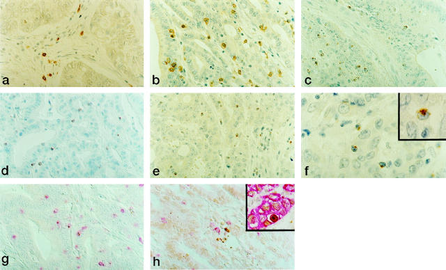
Microsatellite instability (MSI) characterizes colorectal carcinomas (CRCs) in hereditary nonpolyposis colorectal cancer (HNPCC) syndrome and a proportion of sporadic CRCs. These MSI+ CRCs share several clinicopathological features, including a reputation for better survival rates than MSI- cases and a pronounced stromal inflammatory reaction of still undefined nature. In the present study, the presence, spatial distribution, and activation status of infiltrating cytotoxic effectors were investigated comparatively in 18 MSI+ and 37 MSI- CRCs by immunohistochemistry. The frequency of apoptosis was also evaluated by morphology and in situ end-labeling. MSI+ cases carried significantly higher numbers of cytotoxic lymphocytes infiltrating within neoplastic epithelial structures, as shown by immunostaining for CD3 (15.1 +/- 6.2 versus 4.6 +/- 4.1, P < 0.001), CD8 (13 +/- 6.4 versus 3.7 +/- 3.8, P < 0.001), and TIA-1 (11.2 +/- 6.5 versus 1.9 +/- 1.7, P < 0.001). These cytotoxic effectors were globally more activated in MSI+ than in MSI- tumors, as revealed by the expression of granzyme B (5.3 +/- 4.5 versus 0.6 +/- 1.3, P < 0.001). In MSI+ CRCs, the number of intraepithelial activated cytotoxic lymphocytes was significantly correlated with the proximal location of the tumor, a poorly differentiated phenotype, and the presence of peritumor lymphoid nodules. Multivariate analysis revealed that MSI was the major determinant of the presence of activated cytotoxic intraepithelial lymphocytes. Moreover, MSI+ CRCs also showed a significantly higher percentage of tumor cells undergoing apoptotic cell death (4.1 +/- 2.1 versus 2.6 +/- 1.1, P < 0.0001, by the TUNEL method), often located in close proximity of activated cytotoxic lymphocytes. These results are consistent with the presence of anti-tumor cytotoxic immune responses in most of MSI+ CRCs, a phenomenon that may at least in part contribute to the survival advantage ascribed to these patients.
Figures
References
-
- Aaltonen LA, Peltomäki P, Leach FS, Sistonen P, Pylkkänen L, Mecklin JP, Järvinen H, Powell SM, Jen J, Hamilton SR, Petersen GM, Kinzler KW, Vogelstein B, de la Chapelle A: Clues to the pathogenesis of familial colorectal cancer. Science 1993, 260:812-816 - PubMed
-
- Wu C, Akiyama Y, Imai K, Miyake S, Nagasaki H, Oto M, Okabe S, Iwama T, Mitamura K, Masumitsu H, Nomizu T, Baba S, Maruyama K, Yuasa Y: DNA alterations in cells from hereditary non-polyposis colorectal cancer patients. Oncogene 1994, 9:991-994 - PubMed
-
- Thibodeau SN, Bren G, Schaid D: Microsatellite instability in cancer of the proximal colon. Science 1993, 260:816-819 - PubMed
-
- Ionov Y, Peinado MA, Malkhosyan S, Shibata D, Perucho M: Ubiquitous somatic mutations in simple repeated sequences reveal a new mechanism for colonic carcinogenesis. Nature 1993, 363:558-561 - PubMed
-
- Lothe RA, Peltomäki P, Meling GI, Aaltonen LA, Nyström Lahti M, Pylkkänen L, Heimdal K, Andersen TI, Moller P, Rognum TO, Fossa SD, Haldorsen T, Langmark F, Brogger A, de la Chapelle A, Borresen AL: Genomic instability in colorectal cancer: relationship to clinicopathological variables and family history. Cancer Res 1993, 53:5849-5852 - PubMed
Publication types
MeSH terms
Substances
LinkOut - more resources
Full Text Sources
Other Literature Sources
Medical
Research Materials
Miscellaneous
