. 1999 Jun;154(6):1923-32.
doi: 10.1016/S0002-9440(10)65450-8.

A nonhuman primate model for the selective elimination of CD8+ lymphocytes using a mouse-human chimeric monoclonal antibody

Affiliations

A nonhuman primate model for the selective elimination of CD8+ lymphocytes using a mouse-human chimeric monoclonal antibody

J E Schmitz et al. Am J Pathol. 1999 Jun.

Abstract

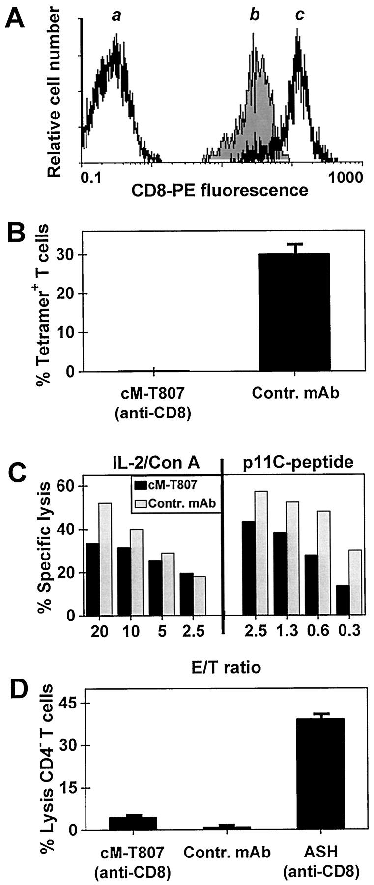
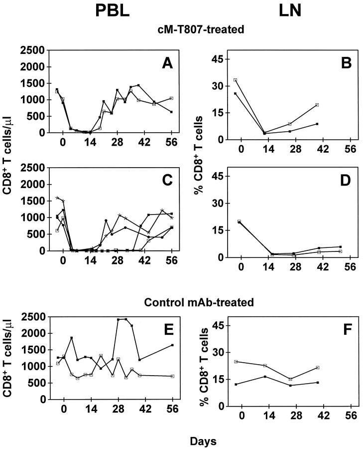
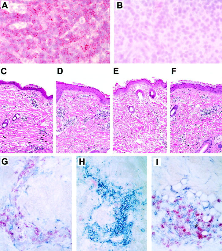
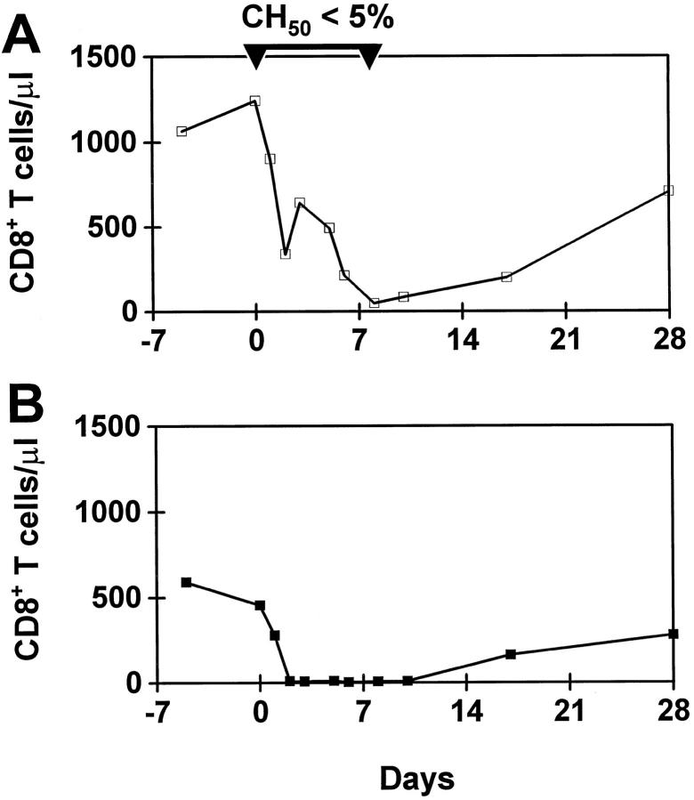
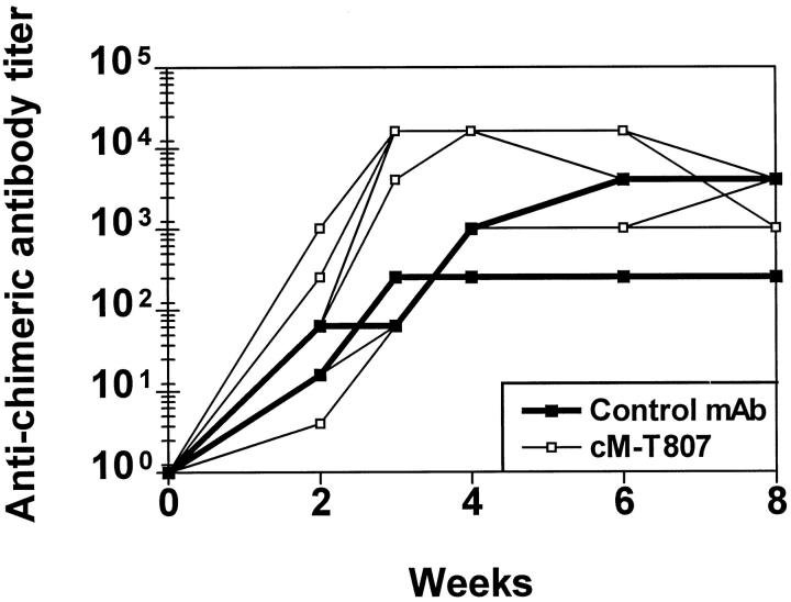
Nonhuman primates provide valuable animal models for human diseases. However, studies assessing the role of cell-mediated immune responses have been difficult to perform in nonhuman primates. We have shown that CD8+ lymphocyte-mediated immunity in rhesus monkeys can be selectively eliminated using the mouse-human chimeric anti-CD8 monoclonal antibody cM-T807. In vitro, this antibody completely blocked antigen-specific expansion of cytotoxic T cells and decreased major histocompatibility complex class I-restricted, antigen-specific lysis of target cells but did not mediate complement-dependent cell lysis. In vivo administration of cM-T807 in rhesus monkeys resulted in near total depletion of CD8+ T cells from the blood and lymph nodes for up to 6 weeks. This depletion was not solely complement-dependent and persisted longer in adults than in juveniles. Preservation of B cell and CD4+ T cell function in monkeys depleted of CD8+ lymphocytes was demonstrated by their ability to develop humoral immune responses to the administered chimeric monoclonal antibody. Furthermore, during CD8+ lymphocyte depletion, monkeys developed delayed-type hypersensitivity reactions comprised only of CD4+ T cells but not CD8+ T cells. This CD8+ lymphocyte depletion model should prove useful in defining the role of cell-mediated immune responses in controlling infectious diseases in nonhuman primates.


Figures

Figure 1.
Figure 1.
Activity of cM-T807 monoclonal antibody on CD8+ lymphocytes in vitro. A: Pretreatment of PBL with saturating concentrations with cM-T807 did not block the detection of CD8+ T cells using anti-CD8-PE. a, CD8- T cells (background staining); b, CD8+ T cells pretreated with cM-T807, then stained with anti-CD8-PE; c, CD8+ T cells stained with anti-CD8-PE without pretreatment. B: cM-T807 blocked the in vitro expansion of viral antigen-specific CD8+ T cells. PBL from a monkey infected with SIVmac were incubated with IL-2 and a MHC class I-restricted SIVmac peptide. Expansion of antigen-specific CD8+ T cells after 7 days of incubation in the presence of cM-T807 or a control monoclonal antibody was determined by the percentage of T cells that bound MHC-I/peptide tetrameric complexes (mean ± SD, n = 3). C: cM-T807 decreased antigen-specific, MHC class I-restricted cell lysis. PBL from 2 SIVmac-infected monkeys were expanded by nonspecific stimulation (Con A/IL-2) or by specific stimulation with a MHC class I-restricted SIVmac peptide (p11C-peptide) and then incubated with SIVmac peptide-pulsed, autologous target cells. Lysis of target cells in the presence of cM-807 (black bars) or control antibody (shaded bars) was determined by chromium release assay. D: cM-T807 caused marginal complement-dependent cell lysis. Human PBL were incubated with complement and cM-T807, control antibody or an anti-CD8 antibody (ASH) known to be lytic. Lysis of CD8+ T cells was determined flow cytometrically by quantitating CD3+4− cells that showed uptake of PI (mean ± SD, n = 3). E/T ratio, effector cell-to-target cell ratio.
Figure 2.
Figure 2.
Intravenous administration of cM-T807 depleted blood and lymph node of CD8+ T cells. Rhesus monkeys received three injections of cM-T807 or control monoclonal antibody (3–5 mg/kg) on days 0, 3, and 7. CD8+ T cells were depleted from the blood (A, C) and lymph nodes (B, D). In monkeys 2 years of age (A, B), duration of depletion was shorter than in monkeys >5 years of age (C, D). The identical treatment protocol with a control monoclonal antibody resulted in no change in CD8+ T cells (E, F). LN, lymph node. Each line illustrates the results from one monkey.
Figure 3.
Figure 3.
Histology and immunohistochemistry of lymph nodes and skin. A-B: cM-T807-treatment resulted in nearly total depletion of CD8+ lymphocytes from lymph nodes as shown in the T-cell zone of lymph nodes. Sections of lymph nodes taken before (A) or 14 days after (B) treatment with cM-T807 were stained for CD8. Original magnification, ×100. C-F: Cellular infiltration in cutaneous DTH reactions in tetanus toxoid-sensitized monkeys. Mononuclear cells infiltrated dermal and subdermal regions of skin at the site of tetanus toxoid injection before treatment (C) and at 2 weeks after cM-T807 treatment when CD8+ lymphocytes were depleted (D). Two to four weeks after the reappearance of CD8+ lymphocytes, saline injection resulted in no cellular infiltrates (E), but tetanus toxoid caused the typical DTH reaction (F). G-I: Immunophenotype of cells infiltrating DTH reaction. Before cM-T807 treatment, both CD4+ (blue) and CD8+ (red) cells appear at the site of the DTH reaction (G). After CD8+ lymphocyte depletion, only CD4+ cells are recruited into the DTH reaction (H), whereas both CD4+ (blue) and CD8+ (red) cells infiltrate skin after reappearance of CD8+ lymphocytes (I). (A, B, immunohistochemistry using New Fuchsin as chromogen; C-E, hematoxylin and eosin staining; G-F, Vector Red and Vector Blue chromogens.)
Figure 4.
Figure 4.
Effect of consumption of complement in vivo through administration of CVF on depletion of CD8+ lymphocytes. A rhesus monkey received 4 injections of CVF (500 μg/kg total dose) to consume complement in vivo during treatment with a single administration of cM-T807 (5 mg/kg, day 0). Although hemolytic complement was reduced to <5% by this treatment, cM-T807 caused substantial depletion of CD8+ T cells (A) as compared to the identical treatment of a control monkey with cM-T807 that did not receive CVF (B).
Figure 5.
Figure 5.
Development of humoral immune responses to cM-T807 or control chimeric antibody in monkeys. Detection of monkey antibodies to mouse-human chimeric antibodies was performed by enzyme-linked immunosorbent assay. All monkeys that received either cM-T807 (open symbols) or control chimeric monoclonal antibody (closed symbols) developed humoral immune responses to the administered antibodies, peaking within 3–6 weeks.

References

    1. Yap KL, Ada GL, McKenzie IFC: Transfer of specific cytotoxic T lymphocytes protects mice inoculated with influenza virus. Nature 1978, 273:238-239 - PubMed
    1. Riddell SR, Greenberg PD: Principles for adoptive T cell therapy of human viral diseases. Annu Rev Immunol 1995, 13:545-586 - PubMed
    1. Fung-Leung W-P, Schilham MW, Rahemtulla A, Kundig TM, Vollenweider M, Potter J, van Ewijk W, Mak TW: CD8 is needed for development of cytotoxic T cells but not helper T cells. Cell 1991, 65:443-449 - PubMed
    1. Nesic D, Vukmanovic S: MHC class I is required for peripheral accumulation of CD8+ thymic emigrants. J Immunol 1998, 160:3705-3712 - PubMed
    1. Mielke ME, Niedobitek G, Stein H, Hahn H: Acquired resistance to Listeria monocytogenes in mediated by Lyt-2+ T cells independently of the influx of monocytes into granulomatous lesions. J Exp Med 1989, 170:589-594 - PMC - PubMed

Publication types

MeSH terms