. 1999 Jun 1;13(11):1422-37.
doi: 10.1101/gad.13.11.1422.

Regulation of 4E-BP1 phosphorylation: a novel two-step mechanism

Affiliations

Regulation of 4E-BP1 phosphorylation: a novel two-step mechanism

A C Gingras et al. Genes Dev. .

Abstract

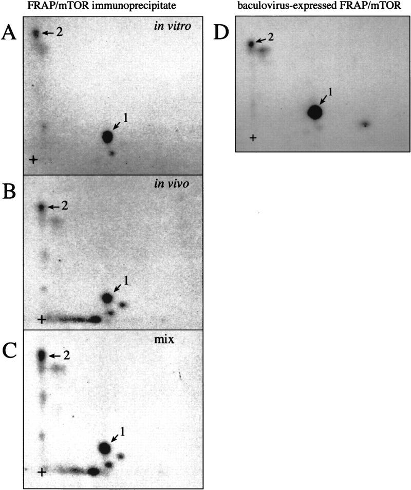
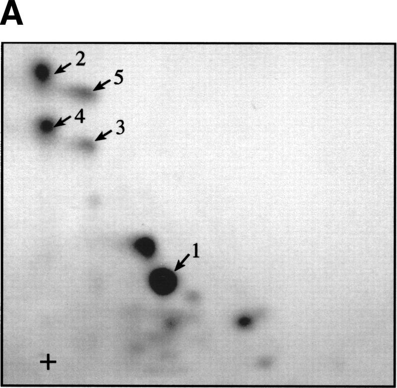
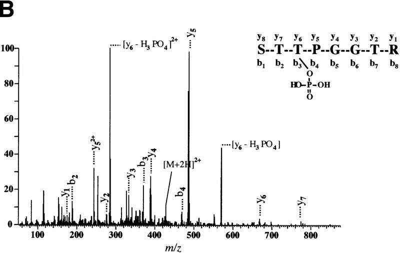
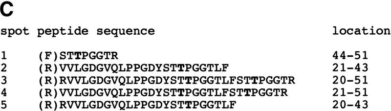
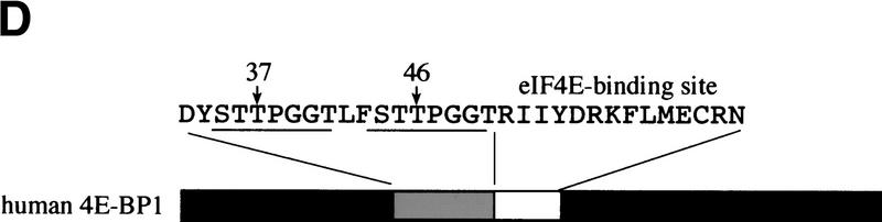
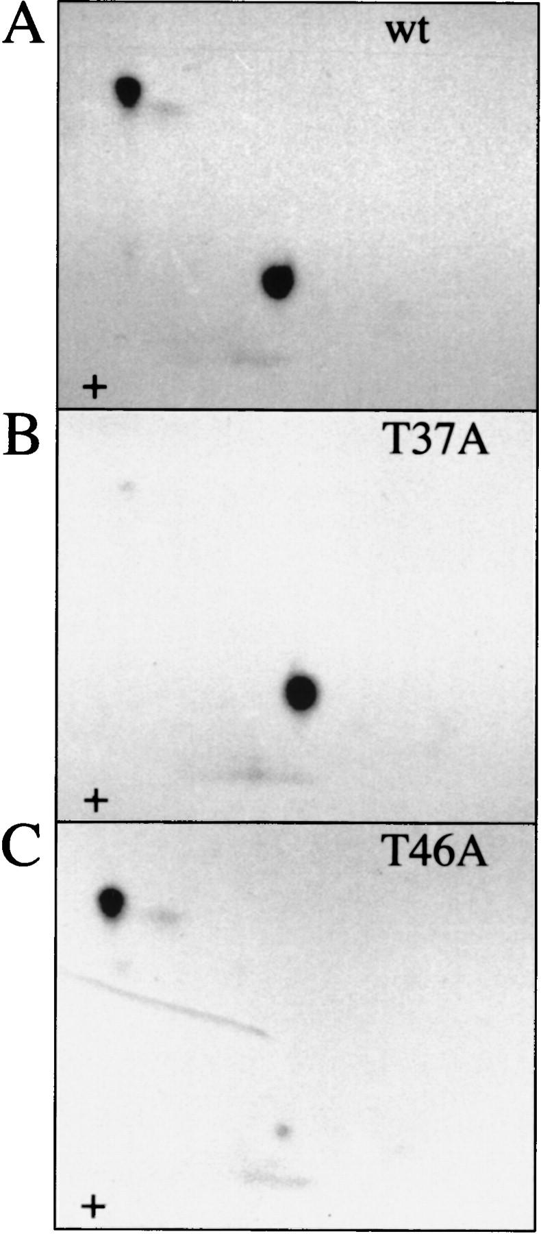
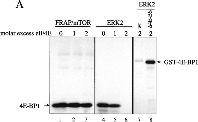
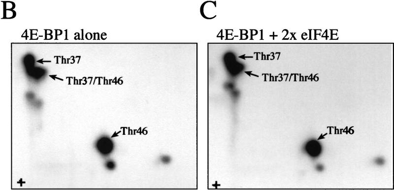
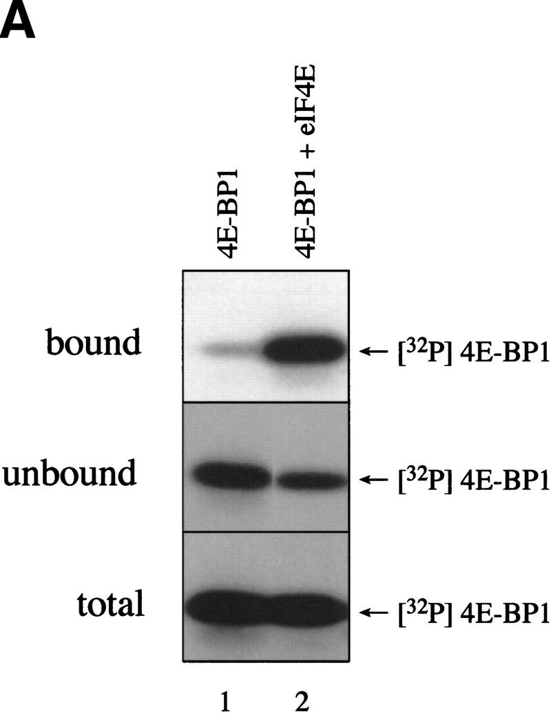
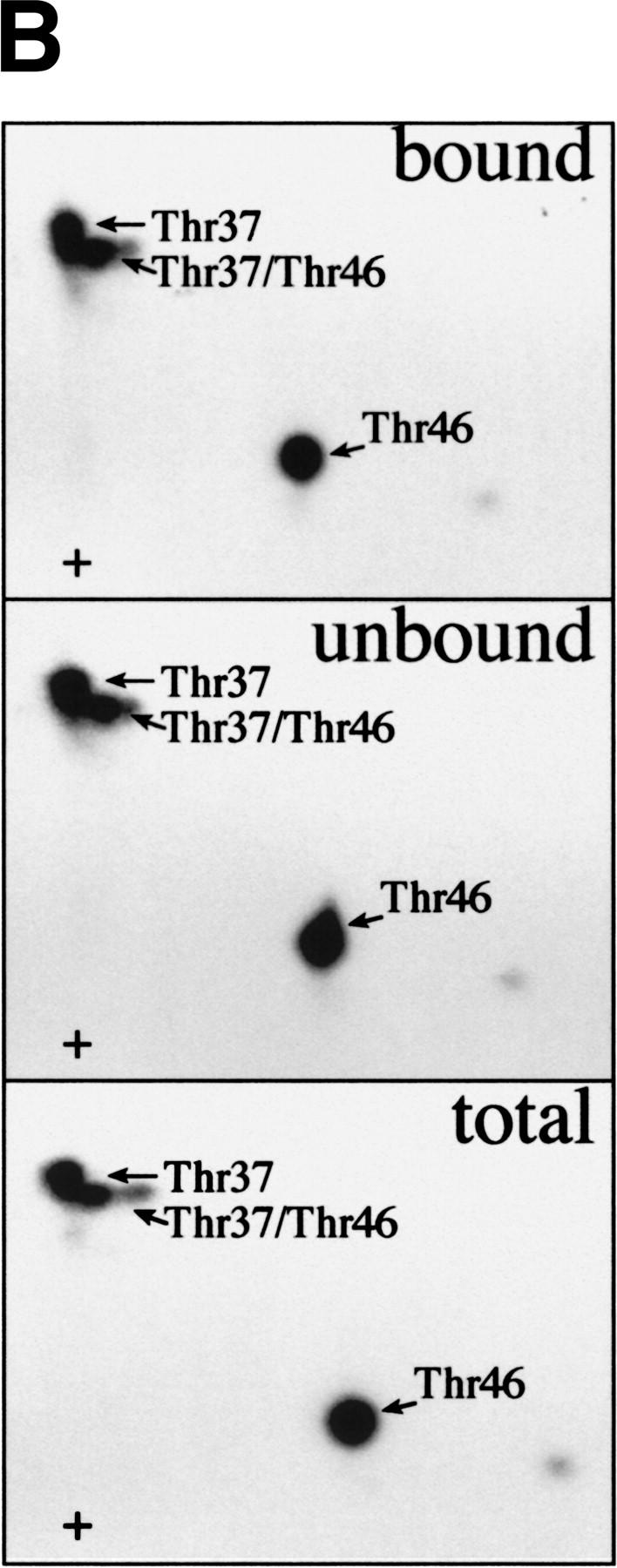
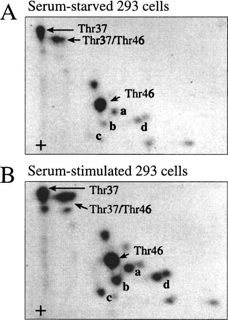
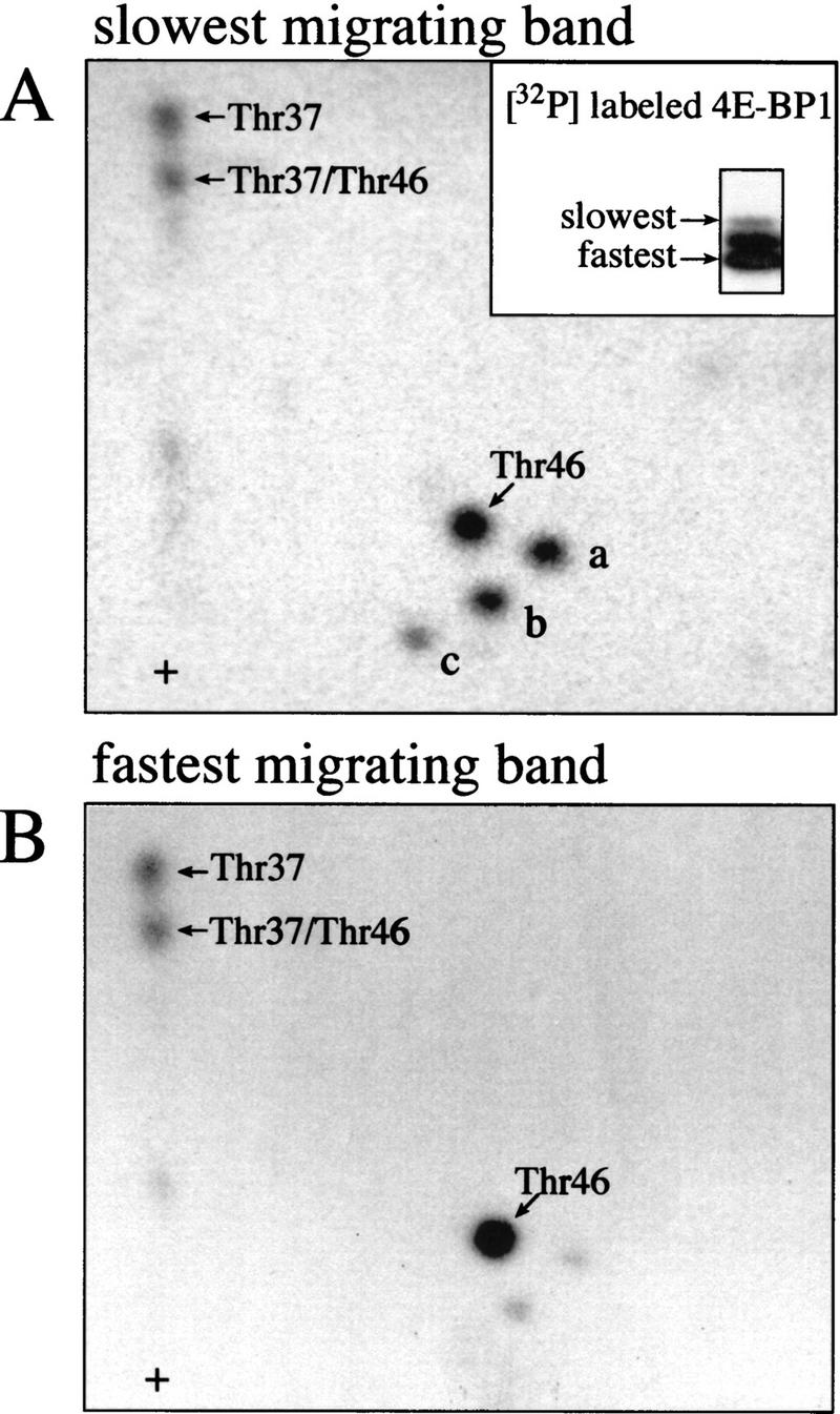
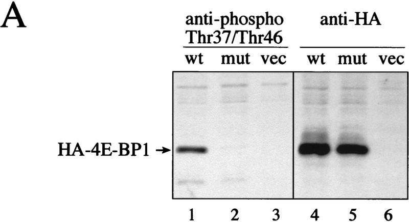
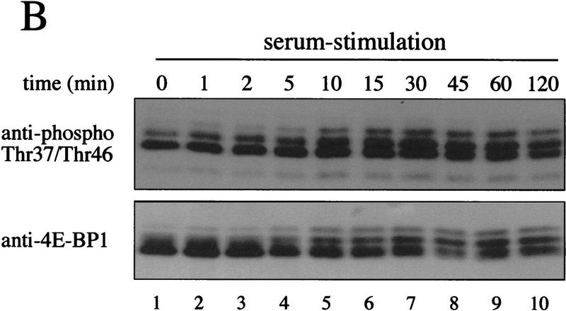
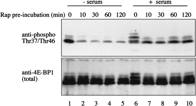
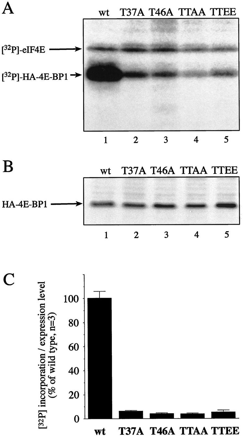
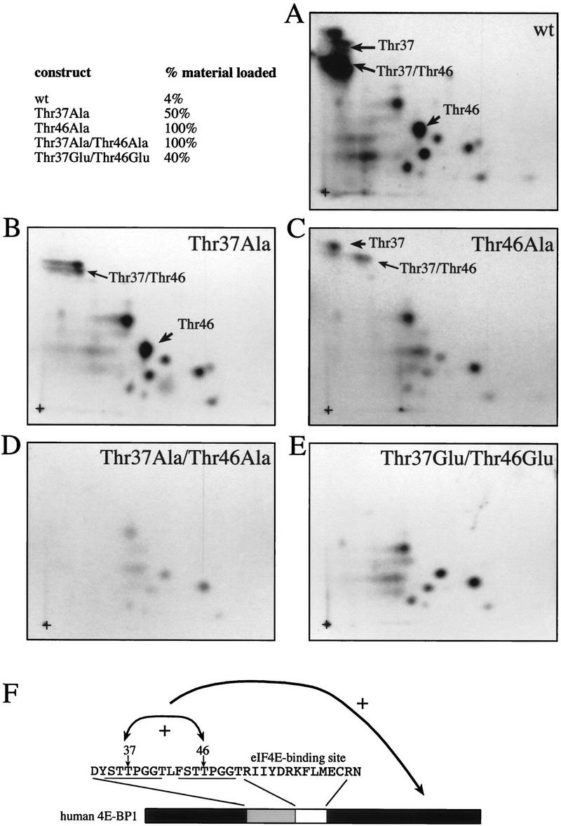
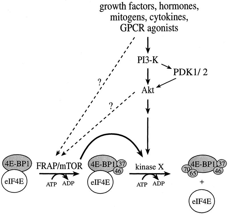
The multisubunit eukaryotic translation initiation factor (eIF) 4F recruits 40S ribosomal subunits to the 5' end of mRNA. The eIF4F subunit eIF4E interacts directly with the mRNA 5' cap structure. Assembly of the eIF4F complex is inhibited by a family of repressor polypeptides, the eIF4E-binding proteins (4E-BPs). Binding of the 4E-BPs to eIF4E is regulated by phosphorylation: Hypophosphorylated 4E-BP isoforms interact strongly with eIF4E, whereas hyperphosphorylated isoforms do not. 4E-BP1 is hypophosphorylated in quiescent cells, but is hyperphosphorylated on multiple sites following exposure to a variety of extracellular stimuli. The PI3-kinase/Akt pathway and the kinase FRAP/mTOR signal to 4E-BP1. FRAP/mTOR has been reported to phosphorylate 4E-BP1 directly in vitro. However, it is not known if FRAP/mTOR is responsible for the phosphorylation of all 4E-BP1 sites, nor which sites must be phosphorylated to release 4E-BP1 from eIF4E. To address these questions, a recombinant FRAP/mTOR protein and a FRAP/mTOR immunoprecipitate were utilized in in vitro kinase assays to phosphorylate 4E-BP1. Phosphopeptide mapping of the in vitro-labeled protein yielded two 4E-BP1 phosphopeptides that comigrated with phosphopeptides produced in vivo. Mass spectrometry analysis indicated that these peptides contain phosphorylated Thr-37 and Thr-46. Thr-37 and Thr-46 are efficiently phosphorylated in vitro by FRAP/mTOR when 4E-BP1 is bound to eIF4E. However, phosphorylation at these sites was not associated with a loss of eIF4E binding. Phosphorylated Thr-37 and Thr-46 are detected in all phosphorylated in vivo 4E-BP1 isoforms, including those that interact with eIF4E. Finally, mutational analysis demonstrated that phosphorylation of Thr-37/Thr-46 is required for subsequent phosphorylation of several carboxy-terminal serum-sensitive sites. Taken together, our results suggest that 4E-BP1 phosphorylation by FRAP/mTOR on Thr-37 and Thr-46 is a priming event for subsequent phosphorylation of the carboxy-terminal serum-sensitive sites.


Figures

Figure 1
Figure 1
FRAP/mTOR phosphorylates 4E-BP1 in vitro on two sites that are also phosphorylated in vivo. (A) FRAP/mTOR immunoprecipitated from rat brain was utilized in an in vitro kinase assay with human 4E-BP1 (expressed as a GST fusion protein, which was then cleaved and HPLC purified) as a substrate. Phosphopeptide mapping was performed on phosphorylated 4E-BP1. The two most intense phosphopeptides are numbered 1 and 2. (+) The origin of migration. (B) Tryptic/chymotryptic map of 4E-BP1 phosphorylated in 293 cells. The positions of phosphopeptides that comigrate with the FRAP/mTOR phosphorylated peptides 1 and 2 are indicated. (C) An equal number of cpm from the in vitro- and the in vivo-phosphorylated 4E-BP1 were combined and phosphopeptide mapping was performed. (D) Tryptic/chymotryptic map of 4E-BP1 phosphorylated in vitro with purified FRAP/mTOR produced using the baculovirus system.
Figure 2
Figure 2
Thr-37 and Thr-46 are phosphorylated in vivo on phosphopeptides that comigrate with FRAP/mTOR phosphorylated peptides. 4E-BP1 immunoprecipitated from 2 × 108 cells (1/20 labeled with [32P]orthophosphate) was separated by SDS-15% PAGE, electroblotted onto a nitrocellulose membrane, and proteolytically cleaved with trypsin/chymotrypsin as described in Materials and Methods. (A) Tryptic/chymotryptic two-dimensional phosphopeptide map of 4E-BP1. Phosphopeptides (labeled 1–5) were isolated from the plates and identified by mass spectrometry. (B) Tandem mass spectrum of m/z 428.9 revealing the sequence and phosphorylation site of the phosphopeptide contained in spot 1 of the map shown in A. The position of the phosphorylated threonine residue (Thr-46) is shown. (C) Identification of the phosphorylated peptides in spots 1–5 from the phosphopeptide map shown in A. Phosphorylated amino acids are in boldface type; the amino acid amino terminal to the cleavage site for trypsin or chymotrypsin is indicated in parentheses. (D) The positions of Thr-37 and Thr-46 in 4E-BP1, relative to the eIF4E-binding site.
Figure 2
Figure 2
Thr-37 and Thr-46 are phosphorylated in vivo on phosphopeptides that comigrate with FRAP/mTOR phosphorylated peptides. 4E-BP1 immunoprecipitated from 2 × 108 cells (1/20 labeled with [32P]orthophosphate) was separated by SDS-15% PAGE, electroblotted onto a nitrocellulose membrane, and proteolytically cleaved with trypsin/chymotrypsin as described in Materials and Methods. (A) Tryptic/chymotryptic two-dimensional phosphopeptide map of 4E-BP1. Phosphopeptides (labeled 1–5) were isolated from the plates and identified by mass spectrometry. (B) Tandem mass spectrum of m/z 428.9 revealing the sequence and phosphorylation site of the phosphopeptide contained in spot 1 of the map shown in A. The position of the phosphorylated threonine residue (Thr-46) is shown. (C) Identification of the phosphorylated peptides in spots 1–5 from the phosphopeptide map shown in A. Phosphorylated amino acids are in boldface type; the amino acid amino terminal to the cleavage site for trypsin or chymotrypsin is indicated in parentheses. (D) The positions of Thr-37 and Thr-46 in 4E-BP1, relative to the eIF4E-binding site.
Figure 3
Figure 3
In vitro kinase assay of Thr-37–Ala and Thr-46–Ala mutant proteins confirm that they are the primary FRAP/mTOR phosphorylation sites. Histidine-tagged murine 4E-BP1 was phosphorylated in vitro in an immune-complex kinase assay with FRAP/mTOR, and tryptic/chymotryptic maps were performed. (A) Wild type; (B) Thr-37–Ala; (C) Thr-46–Ala.
Figure 4
Figure 4
FRAP/mTOR phosphorylates 4E-BP1 complexed with eIF4E. (A) 4E-BP1 was pre-incubated without eIF4E, with an equimolar quantity or with a twofold molar excess of eIF4E, and subjected to a kinase assay using FRAP/mTOR immunoprecipitated from rat brain (lanes 1–3) or recombinant ERK2 (lanes 4–8). Phosphorylated proteins were separated via SDS-15%-PAGE, transferred to nitrocellulose membranes, and subjected to autoradiography. The substrates used were as follows. (Lanes 1–6) A thrombin-cleaved and HPLC-purified GST–4E-BP1; (lane 7) an uncleaved GST–4E-BP1 wild-type protein; (lane 8) an uncleaved GST–4E-BP1 mutant lacking the eIF4E-binding site. (B) Tryptic/chymotryptic map of 4E-BP1 alone (A, lane 1). (C) Tryptic/chymotryptic map of 4E-BP1 complexed with a twofold molar excess eIF4E (A, lane 3).
Figure 4
Figure 4
FRAP/mTOR phosphorylates 4E-BP1 complexed with eIF4E. (A) 4E-BP1 was pre-incubated without eIF4E, with an equimolar quantity or with a twofold molar excess of eIF4E, and subjected to a kinase assay using FRAP/mTOR immunoprecipitated from rat brain (lanes 1–3) or recombinant ERK2 (lanes 4–8). Phosphorylated proteins were separated via SDS-15%-PAGE, transferred to nitrocellulose membranes, and subjected to autoradiography. The substrates used were as follows. (Lanes 1–6) A thrombin-cleaved and HPLC-purified GST–4E-BP1; (lane 7) an uncleaved GST–4E-BP1 wild-type protein; (lane 8) an uncleaved GST–4E-BP1 mutant lacking the eIF4E-binding site. (B) Tryptic/chymotryptic map of 4E-BP1 alone (A, lane 1). (C) Tryptic/chymotryptic map of 4E-BP1 complexed with a twofold molar excess eIF4E (A, lane 3).
Figure 5
Figure 5
FRAP/mTOR phosphorylation of Thr-37 and Thr-46 does not disrupt the 4E-BP1/eIF4E complex. (A) 4E-BP1 alone, or a 4E-BP1/eIF4E complex, was incubated with FRAP/mTOR, as in Fig. 4. The kinase reaction was stopped and one-half of the material was immediately subjected to SDS-PAGE (bottom). The remaining material was incubated with m7GDP-agarose beads to purify eIF4E and associated proteins. The bound (top) and unbound (middle) fractions were analyzed by SDS-PAGE. (B) Phosphorylated 4E-BP1 (A, lane 2; bound, unbound, and total) was analyzed by two-dimensional phosphopeptide mapping.
Figure 5
Figure 5
FRAP/mTOR phosphorylation of Thr-37 and Thr-46 does not disrupt the 4E-BP1/eIF4E complex. (A) 4E-BP1 alone, or a 4E-BP1/eIF4E complex, was incubated with FRAP/mTOR, as in Fig. 4. The kinase reaction was stopped and one-half of the material was immediately subjected to SDS-PAGE (bottom). The remaining material was incubated with m7GDP-agarose beads to purify eIF4E and associated proteins. The bound (top) and unbound (middle) fractions were analyzed by SDS-PAGE. (B) Phosphorylated 4E-BP1 (A, lane 2; bound, unbound, and total) was analyzed by two-dimensional phosphopeptide mapping.
Figure 6
Figure 6
Phosphorylated Thr-37 and Thr-46 are present at a high stoichiometry in serum-starved 293 cells. 293 cells were grown to confluence and serum starved for 30 hr. 32P-Labeling was performed for 4 hr and phosphopeptide maps were generated from starved cells (A) or cells stimulated for 30 min with 10% dialyzed FCS (B). Phosphopeptides containing phospho-Thr-37 or -Thr-46 are identified. Serum-induced phosphopeptides are labeled a–d.
Figure 7
Figure 7
Phosphorylated Thr-37 and Thr-46 are present in the fastest migrating phosphorylated 4E-BP1 isoform. 293 cells were serum starved for 30 hr, incubated with [32P]orthophosphate, stimulated with 10% dialyzed FCS, and extracts were subjected to SDS-PAGE and autoradiography (A, inset). The slowest (A) and fastest-migrating (B) 4E-BP1 isoforms were isolated and phosphopeptide maps were performed.
Figure 8
Figure 8
Phosphospecific antibodies confirm the relative lack of serum sensitivity of Thr-37 and Thr-46. (A) 293 cells were transfected with wild-type HA–4E-BP1 (wt), HA–4E-BP1 Thr-37–Ala/Thr-46–Ala (mut), or vector alone (vec). Cell extracts were prepared and analyzed by Western blotting with either the phosphospecific antibody to Thr-37 and Thr-46 or with an anti-HA antibody. (B) A time course of serum stimulation (after 30 hr of starvation) of 293 cells was performed for the indicated times. Extracts were prepared for Western blotting (see Materials and Methods) and duplicate blots were performed. Phosphospecific antibody to Thr-37 and Thr-46 (see Materials and Methods; top) and an anti-4E-BP1 antibody raised against the entire protein (bottom) were used to probe the membranes.
Figure 8
Figure 8
Phosphospecific antibodies confirm the relative lack of serum sensitivity of Thr-37 and Thr-46. (A) 293 cells were transfected with wild-type HA–4E-BP1 (wt), HA–4E-BP1 Thr-37–Ala/Thr-46–Ala (mut), or vector alone (vec). Cell extracts were prepared and analyzed by Western blotting with either the phosphospecific antibody to Thr-37 and Thr-46 or with an anti-HA antibody. (B) A time course of serum stimulation (after 30 hr of starvation) of 293 cells was performed for the indicated times. Extracts were prepared for Western blotting (see Materials and Methods) and duplicate blots were performed. Phosphospecific antibody to Thr-37 and Thr-46 (see Materials and Methods; top) and an anti-4E-BP1 antibody raised against the entire protein (bottom) were used to probe the membranes.
Figure 9
Figure 9
Sensitivity of Thr-37 and Thr-46 phosphorylation to rapamycin. Serum-starved 293 cells were incubated with rapamycin (20 ng/ml). One set of plates was lysed immediately (lanes 1–5); the other was serum stimulated for 30 min prior to lysis (lanes 6–10). Extracts were heat treated and analyzed by Western blotting, as described in Materials and Methods. The membranes were incubated with either an anti-phosphospecific antibody (top) or with an anti-4E-BP1 antibody (bottom).
Figure 10
Figure 10
Mutation of Thr-37 or Thr-46 drastically reduces 4E-BP1 phosphorylation in vivo. 4E-BP1 wild type (wt) and mutants Thr-37–Ala (T37A), Thr-46–Ala (T46A), Thr-37–Ala/Thr-46–Ala (TTAA), and Thr-37–Glu/Thr-46–Glu (TTEE) in a pcDNA3-3HA vector were transfected into 293T cells (six plates/construct). Three transfected plates were subjected to 32P-labeling, and HA-tagged 4E-BP1 was immunoprecipitated using an anti-HA antibody (HA.11). Precipitated proteins were subjected to SDS-PAGE, transferred to nitrocellulose membranes, and autoradiographed. The intensity of the HA–4E-BP1 bands was quantified by PhosphorImager. The three other plates were used to analyze the expression levels of the HA-tagged proteins by Western blotting, using anti-HA as the primary antibody and 125I-coupled anti-mouse IgG as the secondary antibody. Expression level was quantified by PhosphorImager. The average of the 32P incorporation into the mutant proteins was normalized to the expression level of each mutant. (A) A representative autoradiograph is shown; arrows indicate 32P-labeled HA–4E-BP1 and coprecipitated eIF4E. (B) A representative Western blot showing the levels of the mutants; this Western blot was developed by chemiluminescence, which produces a brighter signal. (C) Incorporation of 32P into each mutant, normalized to the expression level. The incorporation in the wild-type protein is set to 100%.
Figure 11
Figure 11
Effects of mutations of Thr-37 and/or Thr-46 on other phosphorylation sites. (A–E) Phosphopeptide maps were performed on HA-tagged 4E-BP1 proteins from Fig. 10. (F) Thr-37 and Thr-46 affect 4E-BP1 phosphorylation. The arrows indicate (1) a mutually positive effect of Thr-37 and Thr-46 phosphorylation, and (2) a positive effect of Thr-37 and Thr-46 phosphorylation on the phosphorylation of the serum-sensitive sites. The fraction of samples from Fig. 10 loaded onto each map is shown (upper left). An equivalent quantity in cpm was loaded for each mutant.
Figure 12
Figure 12
A two-step model for the phosphorylation of 4E-BP1. FRAP/mTOR phosphorylates 4E-BP1 (in a 4E-BP1/eIF4E complex) on Thr-37 and Thr-46. Phosphorylation of Thr-37 and Thr-46 is a prerequisite for an unidentified serum-sensitive kinase (acting downstream of Akt) to phosphorylate 4E-BP1 on the serum-sensitive sites. The latter phosphorylation triggers the release of 4E-BP1 from eIF4E. A direct link between Akt and FRAP/mTOR remains to be proven.

References

    1. Banin S, Moyal L, Shieh S, Taya Y, Anderson CW, Chessa L, Smorodinsky NI, Prives C, Reiss Y, Shiloh Y, Ziv Y. Enhanced phosphorylation of p53 by ATM in response to DNA damage. Science. 1998;281:1674–1677. - PubMed
    1. Beretta L, Svitkin YV, Sonenberg N. Rapamycin stimulates viral protein synthesis and augments the shutoff of host protein synthesis upon picornavirus infection. J Virol. 1996;70:8993–8996. - PMC - PubMed
    1. Brooks RF. Continuous protein synthesis is required to maintain the probability of entry into S phase. Cell. 1977;12:311–317. - PubMed
    1. Brown EJ, Beal PA, Keith CT, Chen J, Shin TB, Schreiber SL. Control of p70 s6 kinase by kinase activity of FRAP in vivo. Nature. 1995;377:441–446. - PubMed
    1. Brunn GJ, Williams J, Sabers C, Wiederrecht G, Lawrence JC, Jr, Abraham RT. Direct inhibition of the signaling functions of the mammalian target of rapamycin by the phosphoinositide 3-kinase inhibitors, wortmannin and LY294002. EMBO J. 1996;15:5256–5267. - PMC - PubMed

Publication types

MeSH terms

Substances