. 1999 Jun 1;13(11):1438-52.
doi: 10.1101/gad.13.11.1438.

A PDK1 homolog is necessary and sufficient to transduce AGE-1 PI3 kinase signals that regulate diapause in Caenorhabditis elegans

Affiliations

A PDK1 homolog is necessary and sufficient to transduce AGE-1 PI3 kinase signals that regulate diapause in Caenorhabditis elegans

S Paradis et al. Genes Dev. .

Abstract

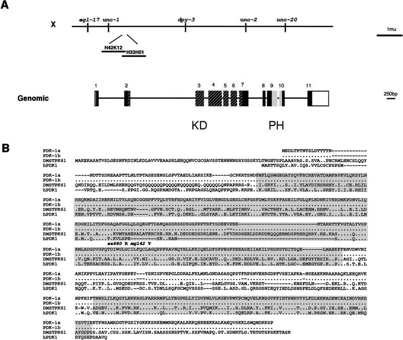
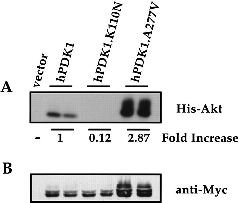
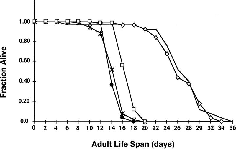
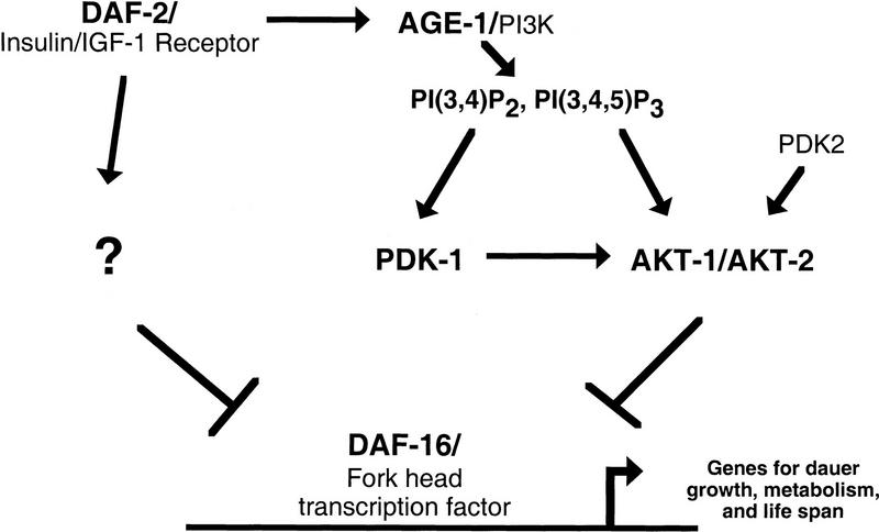
An insulin receptor-like signaling pathway regulates Caenorhabditis elegans metabolism, development, and longevity. Inactivation of the insulin receptor homolog DAF-2, the AGE-1 PI3K, or the AKT-1 and AKT-2 kinases causes a developmental arrest at the dauer stage. A null mutation in the daf-16 Fork head transcription factor alleviates the requirement for signaling through this pathway. We show here that a loss-of-function mutation in pdk-1, the C. elegans homolog of the mammalian Akt/PKB kinase PDK1, results in constitutive arrest at the dauer stage and increased life span; these phenotypes are suppressed by a loss of function mutation in daf-16. An activating mutation in pdk-1 or overexpression of wild-type pdk-1 relieves the requirement for AGE-1 PI3K signaling. Therefore, pdk-1 activity is both necessary and sufficient to propagate AGE-1 PI3K signals in the DAF-2 insulin receptor-like signaling pathway. The activating mutation in pdk-1 requires akt-1 and akt-2 gene activity in order to suppress the dauer arrest phenotype of age-1. This indicates that the major function of C. elegans PDK1 is to transduce signals from AGE-1 to AKT-1 and AKT-2. The activating pdk-1 mutation is located in a conserved region of the kinase domain; the equivalent amino acid substitution in human PDK1 activates its kinase activity toward mammalian Akt/PKB.


Figures

Figure 1
Figure 1
pdk-1 encodes a serine/threonine kinase. (A, top) Genetic and physical map of the pdk-1 region. pdk-1 is contained on fosmids H42K12 and H33H01. The starting methionine of PDK-1 is at 9848 bp on H42K12, the PDK-1 stop codon is at 3082 bp on H33H01; the fosmids overlap for 1201 bp (from 12897 to end on fosmid H42K12). (Bottom) Exon/intron structure of pdk-1. Coding regions are solid boxes; noncoding regions are open boxes; introns are lines. The kinase domain is indicated by hatched boxes (Hanks and Hunter 1995); the pleckstrin homology domain is indicated by the shaded boxes (Alessi et al. 1997a). (B) PDK-1a (GenBank accession no. AF130406); PDK1b (no. AF130407), DMSTPK61(EMBL Y07908), and human PDK1(GenBank accession no. AF017995) were aligned using PILEUP (GCG) (accession numbers for the proteins used in the PILEUP are indicated in parentheses). Identical residues are indicated by dots; gaps introduced to align the sequence are indicated by dashes. The kinase domain is indicated by the amino-terminal shaded area and the pleckstrin homology domain is indicated by the carboxy-terminal shaded area. The sa680 Gly-295–Arg substitution is indicated by an R above the PDK1a sequence. The mg142 Ala-303–Val substitution is indicated by a V above the PDK1a sequence.
Figure 2
Figure 2
Substitution of A277V in human PDK1 leads to activation of the enzyme. Wild-type human PDK1 (hPDK1), kinase inactive hPDK1 (hPDK1.K110N), and the activating A277V mutation in hPDK1 (hPDK1.A277V) expression clones were transfected transiently in duplicate into 293T cells and after the transfection protocol, the protein kinase activity of each mutant toward Akt/PKB was assayed in an in vitro protein kinase assay (see Material and Methods). The data are representative of four independent experiments. (A) Relative amounts of hPDK1 proteins (hPDK1, hPDK1.K110N, and hPDK1.A277V) were normalized for hPDK1 protein concentration from B using anti-Myc antibody and ECL detection (Amersham) on a Bio-Rad Molecular Imager, and the adjusted amounts of lysate used for immunoprecipitation with anti-Myc antibody. The immunoprecipitates were washed and an in vitro kinase assay using His–Akt as substrate was carried out. The phosphorylation of Akt/PKB (His–Akt) was assessed by autoradiography and quantitated on a Bio-Rad Molecular Imager. The data are normalized to wild-type hPDK1 activity (1) and represent the average of the two lanes for each hPDK1 construct. In the hPDK1.A277V lanes, only the lower species (comigrating with His–Akt in the wild-type hPDK1 lanes) were used for quantitation as the identity of the higher migrating species is unclear. (B) Total cell lysate was assayed for total protein content (Bio-Rad Protein Assay) and equal amounts of protein loaded on a 7.5% SDS–polyacrylamide gel, and resolved proteins transferred to nitrocellulose and immunoblotted with anti-Myc antibody. Additional anti-Myc immunoreactive bands are observed with the A277V mutant when compared with either wild-type or kinase-inactive proteins.
Figure 3
Figure 3
Adult life span of pdk-1(sa680) mutants. pdk-1(sa680) extends adult life span and a mutation in daf-16 suppresses the life span increase at 25°C. Mean life spans were 17 days for wild type (□, n = 50), 15 days for pdk-1(mg142) (*, n = 50), 15 days for daf-16(m27); pdk-1(sa680) (●, n = 49), 27 days for pdk-1(sa680) (⋄, n = 50), 27 days for sqt-1(sc13) age-1(hx546) (, n = 25). Mean life spans are significantly different (P≤2e−9; see Materials and Methods) from each other except pdk-1(sa680) compared with sqt-1(sc13) age-1(hx546), and daf-16(m27); pdk-1(sa680) compared with pdk-1(mg142). Life spans were determined in parallel for all strains; data is from one representative experiment that has been performed at least one other time.
Figure 4
Figure 4
PDK-1/GFP expression. (A) PDK-1/GFP expression in an L1 animal. Expression in the procorpus and anterior bulb of the pharynx (ant.ph.) is shown; expression in the entire pharynx is also observed. PDK-1/GFP expression in the cell bodies of many neurons in the head and tail is visible; occasional expression in neuronal nuclei is observed. Expression of PDK-1/GFP is also observed in the hypodermal cells (hyp) of the tail. (B) PDK-1/GFP expression in the intestine (int.) and ventral nerve cord (VNC) of an L1 animal. Anterior is to the left, ventral is down; the animal is slightly twisted because of co-injection with the rol-6 marker. Cell bodies and axons of the VNC neurons (arrowheads) clearly express PDK-1/GFP as do the cell bodies of the intestinal cells (arrows); VNC and intestinal nuclei do not appear to express PDK-1/GFP.
Figure 5
Figure 5
Model for regulation of reproductive growth and metabolism by the DAF-2 insulin receptor-like signaling pathway. On DAF-2 activation, AGE-1 generates PtdIns-3,4-P2 and PtdIns-3,4,5-P3 that are required for PDK-1 and AKT-1/AKT-2 activation. Other parallel pathways from DAF-2 are also activated and all signaling converges on DAF-16. See text for details.

References

    1. Alessi DR, Andjelkovic M, Caudwell B, Cron P, Morrice N, Cohen P, Hemmings BA. Mechanism of activation of protein kinase B by insulin and IGF-1. EMBO J. 1996;15:6541–6551. - PMC - PubMed
    1. Alessi DR, Deak M, Casamayor A, Caudwell FB, Morrice N, Norman DG, Gaffney P, Reese CB, MacDougall CN, Harbison D, Ashworth A, Bownes M. 3-Phosphoinositide-dependent protein kinase-1 (PDK1): Structural and functional homology with the Drosophila DSTPK61 kinase. Curr Biol. 1997a;7:776–789. - PubMed
    1. Alessi DR, James SR, Downes CP, Holmes AB, Gaffney PRJ, Reese CB, Cohen P. Characterization of a 3-phosphoinositide-dependent protein kinase which phosphorylates and activates protein kinase Ba. Curr Biol. 1997b;7:261–269. - PubMed
    1. Alessi DR, Kozlowski MT, Weng QP, Morrice N, Avruch J. 3-Phosphoinositide-dependent protein kinase 1 (PDK1) phosphorylates and activates the p70 S6 kinase in vivo and in vitro. Curr Biol. 1998;8:69–81. - PubMed
    1. Anderson KE, Coadwell J, Stephens LR, Hawkins PT. Translocation of PDK1 to the plasma membrane is important in allowing PDK1 to activate protein kinase B. Curr Biol. 1998;8:684–691. - PubMed

Publication types

MeSH terms

Associated data