. 1999 Jun 18;274(25):17725-32.
doi: 10.1074/jbc.274.25.17725.

Targeted expression of SV40 large T-antigen to visceral smooth muscle induces proliferation of contractile smooth muscle cells and results in megacolon

Affiliations

Targeted expression of SV40 large T-antigen to visceral smooth muscle induces proliferation of contractile smooth muscle cells and results in megacolon

B P Herring et al. J Biol Chem. .

Abstract

Many pathological conditions result from the proliferation and de-differentiation of smooth muscle cells leading to impaired contractility of the muscle. Here we show that targeted expression of SV40 large T-antigen to visceral smooth muscle cells in vivo results in increased smooth muscle cell proliferation without de-differentiation or decreased contractility. These data suggest that the de-differentiation and proliferation of smooth muscle cells, seen in many pathological states, may be independently regulated. In the T-antigen transgenic mice the increased smooth muscle cell proliferation results in thickening of the distal colon. Consequently the distal colon becomes hyper-contractile and impedes the flow of digesta through the colon resulting in enlargement of the colon oral to the obstruction. These transgenic mice thus represent a novel model of megacolon that results from increased smooth muscle cell proliferation rather than altered neuronal innervation.


Figures

Fig. 1
Fig. 1. Telokin-T-antigen transgenic mice develop megacolon
Shown are the dissected digestive tracts (from stomach to rectum) from a telokin-T-antigen transgenic animal (TRANSGENE) and a wild type litter mate (WILD TYPE). P indicates the proximal colon. D indicates the distal colon.
Fig. 2
Fig. 2. Megacolon results in an increase in the circumference of the colon
Hematoxylin- and eosin-stained cross-sections through the proximal colon of wild type and telokin-T-antigen transgenic animals are shown. A, low magnification. B, high magnification showing the position of the longitudinal muscle (LM), circular muscle (CM), submucosa (SM), muscularis mucosa (MM), and epithelial cells of the mucosa (M).
Fig. 3
Fig. 3. Colon of telokin-T-antigen transgenic mice contain enteric ganglia
Cross-sections of distal and proximal colon from wild type (WT) and telokin-T-antigen transgenic (TG) animals stained for NADPH diaphorase activity. Positive nerves are stained purple. Several myoenteric ganglia are indicated by the arrows. Scale bar indicates 50 μm.
Fig. 4
Fig. 4. Visceral smooth muscle targeted expression of SV40 large T-antigen induces smooth muscle cell proliferation
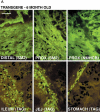
Animals were labeled with tritiated thymidine for 6 h prior to sacrifice. The digestive tract was dissected from the animals, and segments were processed for immunofluorescent analysis as described under “Experimental Procedures.” Cross-sections of gut were stained with antibodies specific for either smooth muscle myosin (SM2) or T-antigen (TAG), both of which are only expressed in smooth muscle cells. Some sections were also stained with antibodies to non-muscle myosin heavy chain B (NMHCB; a generous gift form Dr. Robert Wysolmerski) as indicated. Stained sections were then dipped in photographic emulsion to identify nuclei that had incorporated tritiated thymidine. Staining was visualized under epifluorescence with additional back light illumination to visualize both the antibody staining (green) and the tritiated thymidine-positive nuclei (black granules). Sections shown are from distal colon (DISTAL), proximal colon (PROX), ileum, jejunum (JEJ), and stomach, as indicated. Scale bars represent 50 μm. Representative sections are shown from a 6-month-old telokin-T-antigen transgenic mouse that displayed overt megacolon, from a 6-month-old wild type mouse, and from a 2-month-old telokin-T-antigen transgenic animal that displayed no overt pathology, as indicated.
Fig. 5
Fig. 5. The contractility of colonic muscle from telokin-T-antigen transgenic mice is similar to wild type
Rings of colon (approximately 4 mm in length) were cut from the proximal and distal colon of 2-month-old telokin-T-antigen transgenic (TG, hatched bars) and wild type (WT, solid bars), age-matched animals. Muscles were hung in a myobath, and their maximal contractile responses to various agonists was measured as described under “Experimental Procedures.” A, contractions were initiated by addition of 80 mm KCl. B, contractions were initiated by electrical field stimulation (80 V, 20 Hz, 10 s). C, contractions were initiated by addition of 10–4 m carbachol. Contractions, measured in grams, were normalized to the cross-sectional area of the circular muscle layer in each ring (as this is the muscle layer contributing to the contraction). Three distal and three proximal rings were analyzed from each animal. The data presented represents the mean ± S.E. obtained from 6 animals. Data were statistically analyzed by analysis of variance to identify groups that were statistically different from each other. Statistically significant differences (p < 0.05) are indicated on each graph, together with their p values. Data obtained from wild type animals are indicated by solid bars, and data obtained from transgenic animals are shown by hatched bars. D, the thickness of the circular muscle layer of proximal and distal colon was determined by morphometric analysis of hematoxylin- and eosin-stained sections using Metamorph software. Data represent the mean ± S.E. obtained from a total of at least 15 sections obtained from 4 to 6 different animals. Statistical differences were analyzed by analysis of variance; values having a p value >0.05 were considered not significant. Data obtained from wild type animals is shown by solid bars and from transgenic animals by hatched bars. E and F, dose-response of muscle rings to carbachol. Three distal and three proximal rings were analyzed from each animal. The data presented represent the mean ± S.E. data obtained from 3 animals. Data are expressed as a percentage of the maximal carbachol-induced contraction. E, samples obtained from proximal colon. F, samples obtained from distal colon. Closed symbols indicate data from wild type animals and open symbols from transgenic animals, as indicated.
Fig. 6
Fig. 6. Smooth muscle contractile proteins are not down-regulated in telokin-T-antigen transgenic smooth muscle
Tissue extracts were prepared from samples of proximal (A) and distal (B) colon from 2-month-old wild type and telokin-T-antigen transgenic mice (n = 4). Transgenic mice did not display any overt megacolon. 15 μg of each extract were analyzed by Western blotting with specific antibodies to several contractile protein isoforms. Antibodies used were specific for smooth muscle myosin heavy chain SM1 (SM1), smooth muscle myosin heavy chain SM2 (SM2), non-muscle myosin heavy chain A (nmHCA), non-muscle myosin heavy chain B (nmHCB), h- and l-caldesmon (hCALD and l-CALD), 130-kDa smooth muscle myosin light chain kinase (smMLCK), and 220-kDa non-muscle myosin light chain kinase (nmMLCK), telokin, SM22α, and smooth muscle α-actin. Proteins shown in brackets were not expressed at detected levels in the extracts analyzed. The positions of molecular mass markers are indicated at the left of the blots.

References

    1. Majesky MW, Giachelli CM, Reidy MA, Schwartz SM. Circ. Res. 1992;71:759–768. - PubMed
    1. Ueki N, Sobue K, Kanda K, Hada T, Higashino K. J. Cell Biol. 1987;84:9049–9053. - PMC - PubMed
    1. Glukhova MA, Kabakov AE, Frid MG, Ornatsky OI, Belkin AM, Mukhin DN, Orekhov AN, Koteliansky VE, Smirnov VN. Proc. Natl. Acad. Sci. U. S. A. 1988;85:9542–9546. - PMC - PubMed
    1. Herring BP, Smith AF. Am. J. Physiol. 1996;270:C1656–C1665. - PubMed
    1. Aikawa M, Sivan PN, Kuro-o M, Kimura K, Nakahara K-I, Takewaki S-I, Ueda M, Yamaguchi H, Yazaki Y, Periasamy M, Nagai R. Circ. Res. 1993;73:1002–1012. - PubMed

Publication types

MeSH terms

LinkOut - more resources