. 1999 Jul;73(7):5364-72.
doi: 10.1128/JVI.73.7.5364-5372.1999.

Inhibition of virion maturation by simultaneous deletion of glycoproteins E, I, and M of pseudorabies virus

Affiliations

Inhibition of virion maturation by simultaneous deletion of glycoproteins E, I, and M of pseudorabies virus

A R Brack et al. J Virol. 1999 Jul.

Abstract

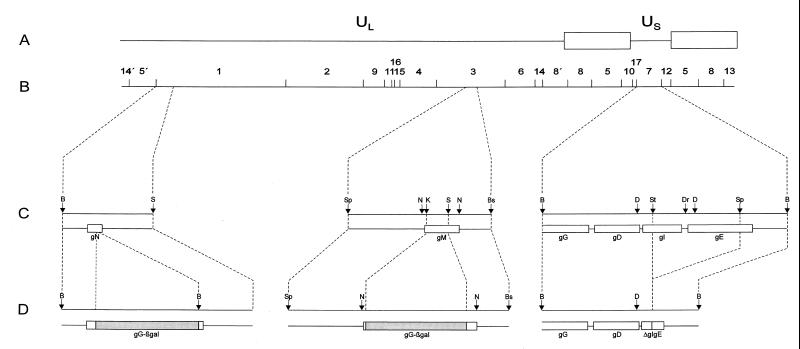
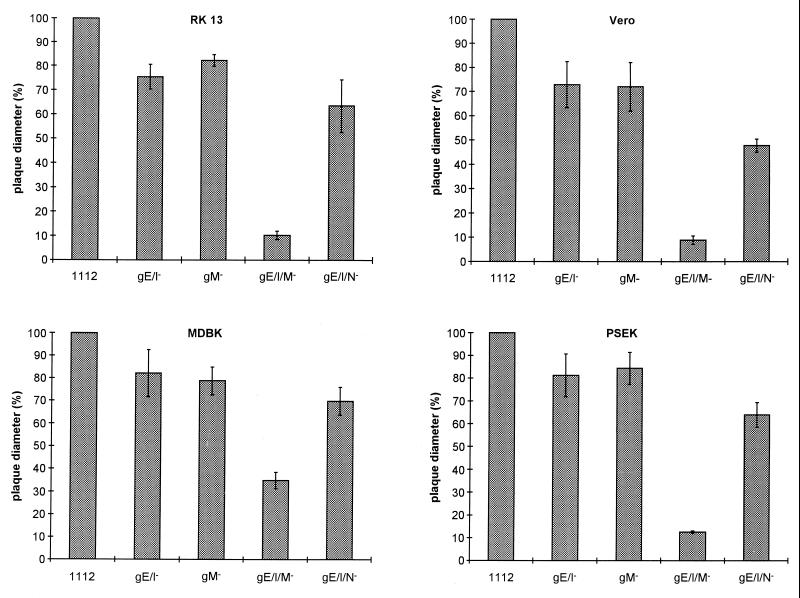
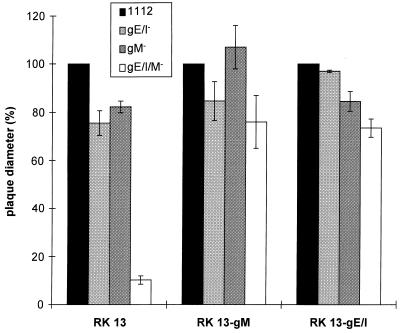
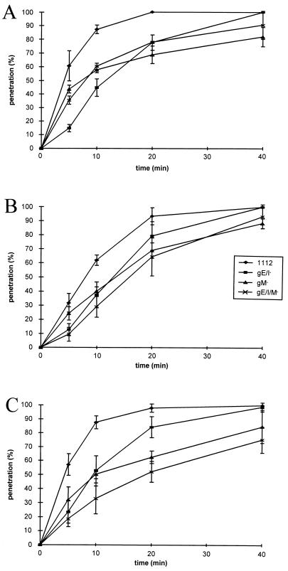
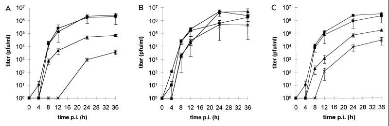
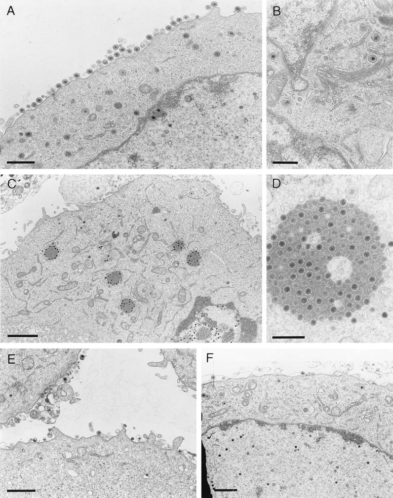
Glycoprotein M (gM), the product of the UL10 gene of pseudorabies virus (PrV), is one of the few nonessential glycoproteins conserved throughout the Herpesviridae. In contrast to wild-type PrV strains, the UL10 gene product of the attenuated PrV vaccine strain Bartha (PrV-Ba) is not modified by N-glycans due to a mutation in the DNA sequence encoding the consensus N-glycosylation motif. To assay function of the UL10 protein in PrV-Ba, a UL10-deletion mutant (PrV-Ba-UL10(-)) was isolated. Surprisingly, in contrast to gM-deleted wild-type PrV, PrV-Ba-UL10(-) was severely impaired in plaque formation, inducing only foci of very few infected RK13, Vero, and PSEK cells and tiny plaques on MDBK cells. Since this effect was significantly more dramatic than in wild-type PrV, additional mutations known to be present in PrV-Ba were analyzed for their contribution to this phenotype. trans-complementation of the mutated PrV-Ba UL21 or gC protein by the wild-type version had no influence on the observed phenotype. In contrast, complementation of the gE/gI deletion rescued the phenotype. The synergistic effect of deletions in gE/gI and gM on plaque size was verified by construction of a gE/I/M triple mutant derived from wild-type PrV which exhibited the same phenotype. The dramatic effect of deletion of gM on plaque size in a gE/I- virus background was mainly attributable to a function of gM, and not of the gM/gN complex, as shown by analysis of a gE/I/N triple mutant. Interestingly, despite the strong effect on plaque size, penetration was not significantly impaired. In noncomplementing cells infected with the gE/I/M triple mutant, electron microscopy showed absence of secondary envelopment in the cytoplasm but occurrence of intracytoplasmic accumulations of nucleocapsids in association with electron dense material, presumably tegument proteins. These structures were not observed after infection of cells expressing either gE/I or gM. We suggest that gE/I and gM are required for late stages in virion morphogenesis prior to final envelopment in the cytoplasm.


Figures

FIG. 1
FIG. 1
Mutations introduced into PrV. (A) Diagram of the PrV genome. It is divided into a unique long (UL) and unique short (US) region by inverted repeats (open rectangles) which bracket the latter. (B) BamHI restriction fragment map. (C) Locations of the glycoprotein genes which had been mutated as demonstrated in panel D. Relevant restriction sites: B, BamHI; Bs, BstEII; D, DdeI; Dr, DraI; N, NlaIII; K, KpnI; S, SalI; Sp, SphI; St, StuI.
FIG. 2
FIG. 2
Plaque morphology of mutants of PrV-Ba. The cell lines indicated on the left were infected under plaque assay conditions with either gG–β-Gal recombinant B80 or UL10–β-Gal recombinant Ba-UL10. Two days after infection, cells were fixed and stained with X-Gal. Bar = 1 mm.
FIG. 3
FIG. 3
Plaque size of PrV-Ba-UL10 on different complementing cells. The cell lines indicated below the bars were infected under plaque assay conditions with PrV-Ba-UL10. Two days after infection, cells were stained with X-Gal, and plaque diameters were measured microscopically and compared to the average diameter of plaques induced by the gG–β-Gal recombinant PrV-B80, which was taken as 100%. Bars indicate standard deviation.
FIG. 4
FIG. 4
Plaque size of different PrV mutants. RK13, Vero, MDBK, and PSEK cells were infected under plaque assay conditions with gG–β-Gal recombinant PrV-1112, PrV-gE/I, PrV-gM, PrV-gE/I/M, or PrV-gE/I/N. Two days after infection, cells were fixed and stained with X-Gal or crystal violet (for PrV-gE/I). Plaque diameters were measured microscopically. The average diameter of plaques induced by PrV-1112 was counted as 100%, and diameters of plaques induced by the other mutant viruses were calculated accordingly. Bars indicate standard deviation.
FIG. 5
FIG. 5
Rescue of plaque formation by PrV-gE/I/M in transcomplementing cells. The cell lines indicated at the bottom were infected with gG–β-Gal recombinant PrV-1112, PrV-gE/I, PrV-gM, or PrV-gE/I/M. Diameters of plaques were measured 2 days after infection under plaque assay conditions. The average diameter of plaques induced by PrV-1112 was counted as 100%, and diameters of plaques induced by the other mutant viruses were calculated accordingly. Bars indicate standard deviation.
FIG. 6
FIG. 6
Penetration kinetics of PrV mutants. Rates of entry of gG–β-Gal recombinant PrV-1112, PrV-gE/I, PrV-gM, or PrV-gE/I/M into wild-type RK13 (A), Vero (B), or MDBK (C) cells were determined as described elsewhere (37). Average values and standard deviation from three independent experiments are depicted.
FIG. 7
FIG. 7
One-step growth analysis of PrV-1112 (⧫), PrV-gE/I (■), PrV-gM (▴), or PrV-gE/I/M (X) in nontransfected (A), gM-expressing (B), or gE/I-expressing (C) RK13 cells. At the indicated times after infection, supernatant and cells were harvested and titrated on RK13 cells, and titers were summed. Average values and standard deviation of two independent experiments are shown.
FIG. 8
FIG. 8
Electron microscopy. Nontransfected RK13 cells were infected with either wild-type PrV (A and B) or PrV-gE/I/M (C and D) and analyzed 16 h after infection. (E and F) RK13-gE/I (E) and RK13-gM (F) cells after infection with the triple mutant. Bars represent 750 nm in panel A, 500 nm in panel B and D, 2 μm in panel C, and 1 μm in panels E and F.

References

    1. Babic N, Klupp B G, Brack A, Mettenleiter T C, Ugolini G, Flamand A. Deletion of glycoprotein gE reduces the propagation of pseudorabies virus in the nervous system of the mouse after intranasal inoculation. Virology. 1996;219:279–284. - PubMed
    1. Baines J D, Roizman B. The UL10 gene of herpes simplex virus 1 encodes a novel viral glycoprotein, gM, which is present in the virion and in the plasma membrane of infected cells. J Virol. 1993;67:1441–1452. - PMC - PubMed
    1. Balan P, Davis-Poynter N, Bell S, Atkinson H, Browne H, Minson T. An analysis of the in vitro and in vivo phenotypes of mutants of herpes simplex virus type 1 lacking glycoproteins gG, gE, gI or the putative gJ. J Gen Virol. 1994;75:1245–1258. - PubMed
    1. Bartha A. Experimental reduction of virulence of Aujeszky’s disease virus. Mag Allatorv Lapja. 1961;16:42–45.
    1. Brack, A., and B. G. Klupp. Unpublished results.

Publication types

Substances

LinkOut - more resources