

Replication, integration, and packaging of plasmid DNA following cotransfection with baculovirus viral DNA
- PMID: 10364295
- PMCID: PMC112604
- DOI: 10.1128/JVI.73.7.5473-5480.1999
Replication, integration, and packaging of plasmid DNA following cotransfection with baculovirus viral DNA
Abstract
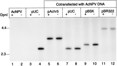
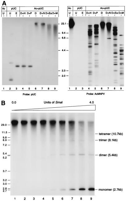
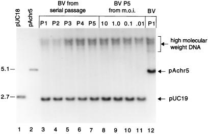
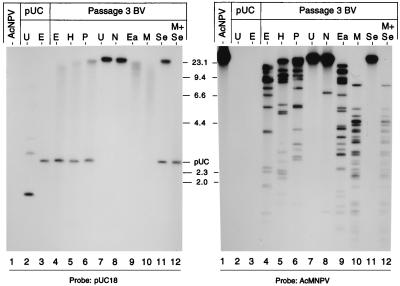
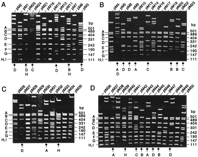
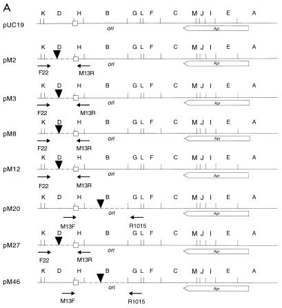
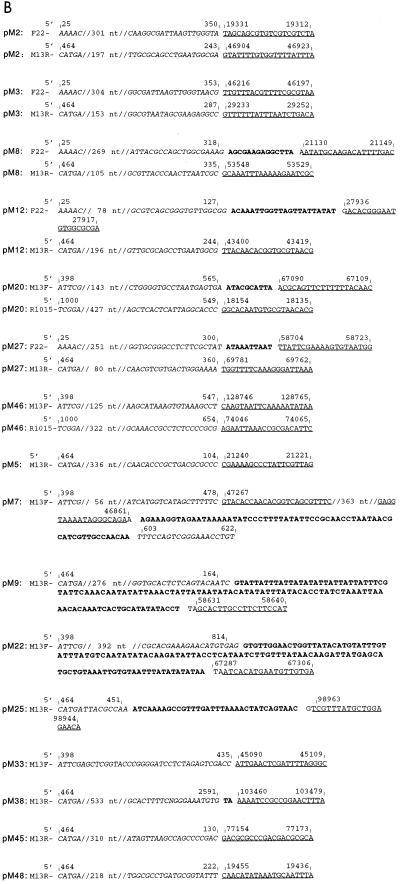
Infection-dependent replication assays have been used to identify numerous putative origins of baculovirus replication. However, plasmid DNA, when cotransfected into insect cells with Autographa californica multinucleocapsid nucleopolyhedrovirus (AcMNPV) DNA, replicates independently of any viral sequence in cis (11). Cotransfection of transfer plasmids and baculovirus DNA is a common procedure used in generating recombinant viruses and in measuring the level of gene expression in transient-expression assays. We have examined the fate of a series of vector plasmids in cotransfection experiments. The data reveal that these plasmids replicate following cotransfection and the replication of plasmid DNA is not due to acquisition of viral putative origin sequences. The conformation of plasmid DNA replicating in the cotransfected cells was analyzed and found to exist as high-molecular-weight concatemers. Ten to 25% of the replicated plasmid DNA was integrated into multiple locations on the viral genome and was present in progeny virions following serial passage. Sequence analysis of plasmid-viral DNA junction sites revealed no homologous or conserved sequences in the proximity of the integration sites, suggesting that nonhomologous recombination was involved during the integration process. These data suggest that while a rolling-circle mechanism could be used for baculovirus DNA replication, recombination may also be involved in this process. Plasmid integration may generate large deletions of the viral genome, suggesting that the process of DNA replication in baculovirus may be prone to generation of defective genomes.
Figures
Similar articles
-
DNA replication promotes high-frequency homologous recombination during Autographa californica multiple nuclear polyhedrosis virus infection.Virology. 1997 Jun 9;232(2):300-9. doi: 10.1006/viro.1997.8573. Virology. 1997. PMID: 9191843
-
No single homologous repeat region is essential for DNA replication of the baculovirus Autographa californica multiple nucleopolyhedrovirus.J Gen Virol. 2007 Jan;88(Pt 1):114-122. doi: 10.1099/vir.0.82384-0. J Gen Virol. 2007. PMID: 17170443
-
Differential requirements for baculovirus late expression factor genes in two cell lines.J Virol. 1995 Oct;69(10):6265-72. doi: 10.1128/JVI.69.10.6265-6272.1995. J Virol. 1995. PMID: 7666527 Free PMC article.
-
DNA-protein interactions during the initiation and termination of plasmid pT181 rolling-circle replication.Prog Nucleic Acid Res Mol Biol. 2003;75:113-37. doi: 10.1016/s0079-6603(03)75004-1. Prog Nucleic Acid Res Mol Biol. 2003. PMID: 14604011 Review.
-
Baculoviruses as gene therapy vectors for human prostate cancer.J Invertebr Pathol. 2011 Jul;107 Suppl:S59-70. doi: 10.1016/j.jip.2011.05.006. J Invertebr Pathol. 2011. PMID: 21784232 Review.
Cited by
-
Identification and Functional Analysis of BmNPV-Interacting Proteins From Bombyx mori (Lepidoptera) Larval Midgut Based on Subcellular Protein Levels.Front Microbiol. 2020 Jun 30;11:1481. doi: 10.3389/fmicb.2020.01481. eCollection 2020. Front Microbiol. 2020. PMID: 32695093 Free PMC article.
-
Differential activity of two non-hr origins during replication of the baculovirus Autographa californica nuclear polyhedrosis virus genome.J Virol. 2000 Jun;74(11):5182-9. doi: 10.1128/jvi.74.11.5182-5189.2000. J Virol. 2000. PMID: 10799593 Free PMC article.
-
Bacteriophage SPP1 Chu is an alkaline exonuclease in the SynExo family of viral two-component recombinases.J Bacteriol. 2003 Apr;185(8):2465-74. doi: 10.1128/JB.185.8.2465-2474.2003. J Bacteriol. 2003. PMID: 12670970 Free PMC article.
-
Oligomerization of Baculovirus LEF-11 Is Involved in Viral DNA Replication.PLoS One. 2015 Dec 14;10(12):e0144930. doi: 10.1371/journal.pone.0144930. eCollection 2015. PLoS One. 2015. PMID: 26660313 Free PMC article.
-
Characterization of the interaction between P143 and LEF-3 from two different baculovirus species: Choristoneura fumiferana nucleopolyhedrovirus LEF-3 can complement Autographa californica nucleopolyhedrovirus LEF-3 in supporting DNA replication.J Virol. 2004 Jan;78(1):329-39. doi: 10.1128/jvi.78.1.329-339.2004. J Virol. 2004. PMID: 14671115 Free PMC article.
References
-
- Ahrens C H, Leisy D J, Rohrmann G F. Baculovirus DNA replication. In: DePamphilis M L, editor. DNA replication in eukaryotic cells. Cold Spring Harbor, New York: Cold Spring Harbor Press; 1996. pp. 855–872.
-
- Ayres M D, Howard S C, Kuzio J, Lopez-Ferber M, Possee R D. The complete DNA sequence of Autographa californica nuclear polyhedrosis virus. Virology. 1994;202:586–605. - PubMed
-
- Bataille D, Epstein A L. Herpes simplex virus type 1 replication and recombination. Biochimie. 1995;77:787–795. - PubMed
-
- Bauser C A, Elick T A, Fraser M J. Characterization of hitchhiker, a transposon insertion frequently associated with baculovirus FP mutants derived upon passage in the TN-368 cell line. Virology. 1996;216:235–237. - PubMed
-
- Carstens E B. Identification and nucleotide sequence of the regions of Autographa californica nuclear polyhedrosis virus genome carrying insertion elements from Spodoptera frugiperda. Virology. 1987;161:8–17. - PubMed
Publication types
MeSH terms
Substances
LinkOut - more resources
Full Text Sources
Other Literature Sources
