. 1999 Jul;73(7):5568-76.
doi: 10.1128/JVI.73.7.5568-5576.1999.

The versatility of paramyxovirus RNA polymerase stuttering

Affiliations

The versatility of paramyxovirus RNA polymerase stuttering

S Hausmann et al. J Virol. 1999 Jul.

Abstract

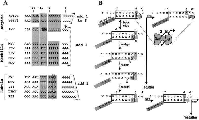
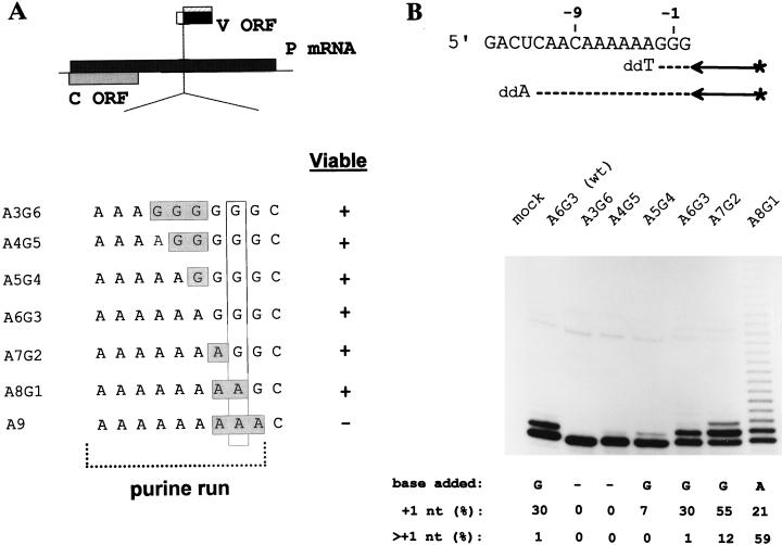
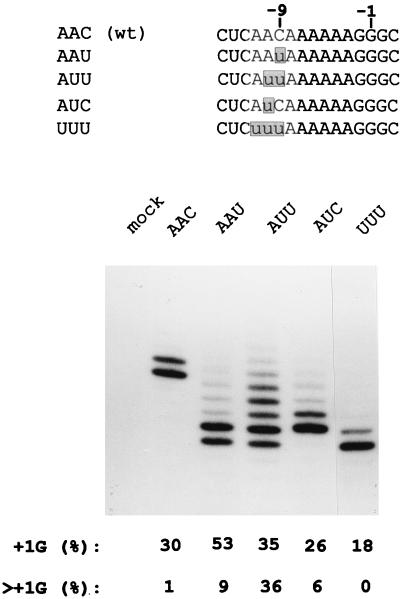
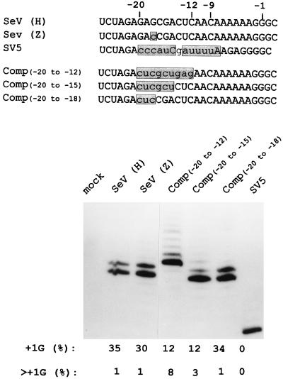
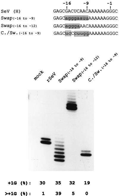
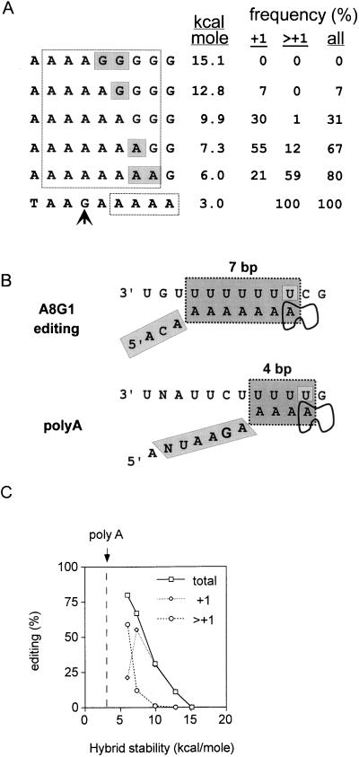
Paramyxoviruses cotranscriptionally edit their P gene mRNAs by expanding the number of Gs of a conserved AnGn run. Different viruses insert different distributions of guanylates, e.g., Sendai virus inserts a single G, whereas parainfluenza virus type 3 inserts one to six Gs. The sequences conserved at the editing site, as well as the experimental evidence, suggest that the insertions occur by a stuttering process, i.e., by pseudotemplated transcription. The number of times the polymerase "stutters" at the editing site before continuing strictly templated elongation is directed by a cis-acting sequence found upstream of the insertions. We have examined the stuttering process during natural virus infections by constructing recombinant Sendai viruses with mutations in their cis-acting sequences. We found that the template stutter site is precisely determined (C1052) and that a relatively short region (approximately 6 nucleotides) just upstream of the AnGn run can modulate the overall frequency of mRNA editing as well as the distribution of the nucleotide insertions. The positions more proximal to the 5' AnGn run are the most important in this respect. We also provide evidence that the stability of the mRNA/template hybrid plays a determining role in the overall frequency and range of mRNA editing. When the template U run is extended all the way to the stutter site, adenylates rather than guanylates are added at the editing site and their distribution begins to resemble the polyadenylation associated with mRNA 3' end formation by the viral polymerase. Our data suggest how paramyxovirus mRNA editing and polyadenylation are related mechanistically and how editing sites may have evolved from poly(A)-termination sites or vice versa.


Figures

FIG. 1
FIG. 1
(A) Sequence homologies at the paramyxovirus editing sites. The sequences are written as [+] RNA, 5′ to 3′, and are grouped into the three genera of the Paramyxovirinae. Spaces have been introduced to emphasize the different elements of the sequence, and shaded boxes indicate sequence conservations. The short G run which is expanded on mRNA editing is shown on the right, together with the pattern of G insertions which occurs for each group. Note that the A run preceding the G run is the only part of this cis-acting sequence that is strictly conserved according to genera. Also note that the second A residue upstream of the rubulavirus G run is replaced by a G (highlighted with a rectangle), which presumably accounts for why rubulaviruses insert a minimum of two G residues when stuttering begins. The precise SeV editing site determined in this study (arrow) is listed as position −1, and positions upstream are numbered according to their distance from this mRNA 3′ end when the polymerase active site is at the editing site. Virus abbreviations: MeV, measles virus; PDV, phocine distemper virus; RPV, rinderpest virus; CDV, canine distemper virus; DMV, dolphin morbillivirus; MuV, mumps virus; PI4, human parainfluenza virus type 4; LPMV, La Piedad, Michoacan virus; PI2, human parainfluenza virus type 2. (b) Competitive kinetic model for SeV RNAP stuttering-elongation decision. The template and mRNA chains of the transcription elongation complex at the editing site are shown schematically. The putative 7-bp hybrid between the polypyrimidine tract of the [−] genome (top strand) and the polypurine run of the nascent mRNA chain (bottom strand) when the transcription elongation complex is at the editing site is boxed. The mRNA upstream of the hybrid is proposed to enter an exit channel (gray-shaded box) before it reaches the surface of the RNAP, which maintains the length of the hybrid as transcription elongation proceeds. The exit channel, analogous to other RNAPs, would contain ∼10 nt (see text). The RNAP bipartite active site, in which the nascent mRNA 3′ end (position −1) and the NTP α-phosphate (position +1) are coordinated via two Mg2+ ions, is highlighted in gray. The transcription complex at the top left is at the editing site (the middle template C1052, boxed in gray) and has just incorporated a strictly templated G1052 (top left). The transcription complex at the editing site presumably pauses due to backsliding of RNAP by one position along both the template and the mRNA chains, undoing the last base pair of the hybrid (and removing the mRNA 3′ end from the active site) and reforming 1 bp on the upstream side. RNAP at pause sites is envisaged as oscillating between the inactive backtracked alignment (second line) and the active alignment (top line). If a strictly templated GMP is the next nucleotide incorporated, RNAP moves past the stutter site and resumes normal elongation (top line). Alternatively, realignment of the hybrid also correctly repositions the mRNA 3′ end in the active site. Hybrid realignment when RNAP is in the backtracked state is initiated when the unpaired 3′ G re-pairs with C−2 (third line), causing the penultimate G to bulge out. Realignment is completed upon translocation of the single nucleotide bulge to the upstream side of the hybrid, reforming a 7-bp hybrid which is nearly as stable as its predecessor. The mRNA 3′ G is now correctly repositioned in the active site, and nucleotide addition at this point leads to a single pseudotemplated G insertion, or stutter (lower case g, bottom line). Having stuttered once, the transcription complex is back to where it started from and has the same choices (second branchpoint, bottom right). Escape from stuttering occurs when the transcription complex moves to a template position where hybrid realignment (stuttering) is no longer favored. Numbers above the genome sequence always indicate the positions relative to mRNA 3′ end at the start of the stutter (top left).
FIG. 2
FIG. 2
mRNA editing in rSeV-A3G6 to -A8G1-infected cells. (A) The ORFs expressed from the P gene mRNA (shaded boxes) are shown above. The sequences of the various rSeVs in which the lengths of the A and G runs were altered (as described in the text) are shown. (B) Parallel cultures of A549 cells were infected with 20 PFU of the various rSeVs per cell as indicated. CsCl pellet RNA was prepared at 24 hpi, and the distribution of lengths of their P gene mRNA purine runs was determined by primer extension analysis limited with ddATP, as schematized in the upper panel. RNA from uninfected cells (mock) served as a negative control. The relative intensities of the various bands was determined, and the fraction of the mRNA population with a single (+1G) or multiple (>+1G) purine insertion is listed below. The lengths of their G runs alone was determined by primer extension analysis limited with ddTTP (not shown), as schematized in the upper panel, and the nature of the insertion deduced (see the text) is also listed below.
FIG. 3
FIG. 3
mRNA editing in rSeV-infected cells with mutations in positions −11 to −9. The various rSeVs with mutations at positions −11 to −9 (relative to the middle G of the G run at position −1) are shown above. The distribution of lengths of their P gene mRNA purine runs determined by primer extension analysis limited with ddATP (as in Fig. 2) are shown below. The lengths of the various extended primers representing the uninserted mRNA (zero bands) vary here due to differences in the position of the limiting ddATP incorporated. The fastest band in each lane represents the zero or uninserted mRNA.
FIG. 4
FIG. 4
mRNA editing in rSeV-infected cells with complementary sequences at positions −20 to −12 and an rSeV with a SV5 editing site. The various rSeVs in which the sequences at positions −20 to −18, −20 to −15, or −20 to −12 were mutated to their complements, as well as one containing positions −20 to −1 of the rubulavirus SV5 sequence (SV5), are shown above. The sequences of SeV strains H and Z, which vary at position −18, are also shown. The distribution of lengths of their P gene mRNA purine runs, as determined by primer extension analysis limited with ddATP (as in Fig. 2 and 3), are shown below.
FIG. 5
FIG. 5
mRNA editing in rSeV-Swap(−16 to −9)-, Swap(−16 to −12)-, and Comp/Swap(−16 to −9)-infected cells. The sequences of the various rSeVs in which positions −16 to −9 were exchanged for those of bPIV3 [Swap(−16 to −9)] or the complement of the bPIV3 sequence [comp/swap(−16 to −9)] or in which the sequences at positions −16 to −12 were exchanged for those of bPIV3 [swap(−16 to −12)] are shown at the top of the figure. The distributions of lengths of their P gene mRNA purine runs, as determined by primer extension analysis limited with ddATP (as in Fig. 2 to 4), are shown below.
FIG. 6
FIG. 6
Importance of hybrid stability in paramyxovirus RNAP stuttering. The 5′ nonapurine runs of the rSeV-AnGn of Fig. 2 are aligned above the conserved SeV poly(A)-termination signal (bottom line of panel A). The seven purines thought to be hybridized to the [−] genome when the transcription elongation complex is at the editing site are boxed, as are the proposed four adenylates hybridized to the [−] genome when the elongation complex is at the polyadenylation site (A). A comparison of the proposed stuttering structures of the rSeV-A8G1 transcription elongation complex at the editing and poly(A)-termination sites is shown in panel B. Note that in the poly(A) structure the stutter site has been displaced downstream by one position relative to the alignment shown in panel A, which would allow for a hybrid with a maximum of only 3 bp. The stabilities of the various hybrids (Serra et al. [49]), along with the distributions of insertions which result, are listed in panel A and are plotted in panel C. The vertical dashed line in panel C indicates the stability of the proposed 4-bp hybrid during polyadenylation as shown in panel B.

References

    1. Barr J N, Whelan S P, Wertz G W. cis-Acting signals involved in termination of vesicular stomatitis virus mRNA synthesis include the conserved AUAC and the U7 signal for polyadenylation. J Virol. 1997;71:8718–8725. - PMC - PubMed
    1. Benne R. RNA editing: an overview. In: Benne R, editor. RNA editing. Chichester, England: Ellis Horwood; 1993. pp. 13–24.
    1. Benne R, Van den Burg J, Brakenhoff J P, Sloof P, Van Boom J H, Tromp M C, Major M C. Transcript of the frameshifted coxII gene from trypanosome mitochondria contains four nucleotides that are not encoded in the DNA. Cell. 1986;46:819–826. - PubMed
    1. Brierley I, Digard P, Inglis S C. Characterization of an efficient coronavirus ribosomal frameshifting signal: requirement for an RNA pseudoknot. Cell. 1989;57:537–547. - PMC - PubMed
    1. Calain P, Roux L. The rule of six: a basic feature for efficient replication of Sendai virus defective interfering RNA. J Virol. 1993;67:4822–4830. - PMC - PubMed

LinkOut - more resources