. 1999 Jul;73(7):5646-53.
doi: 10.1128/JVI.73.7.5646-5653.1999.

Nonhomologous RNA recombination in bovine viral diarrhea virus: molecular characterization of a variety of subgenomic RNAs isolated during an outbreak of fatal mucosal disease

Affiliations

Nonhomologous RNA recombination in bovine viral diarrhea virus: molecular characterization of a variety of subgenomic RNAs isolated during an outbreak of fatal mucosal disease

P Becher et al. J Virol. 1999 Jul.

Abstract

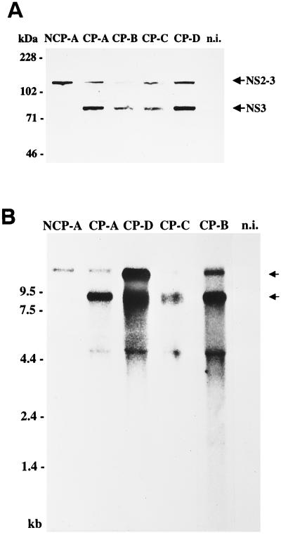
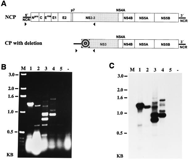
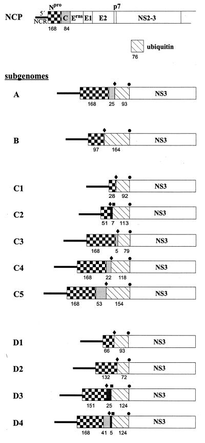
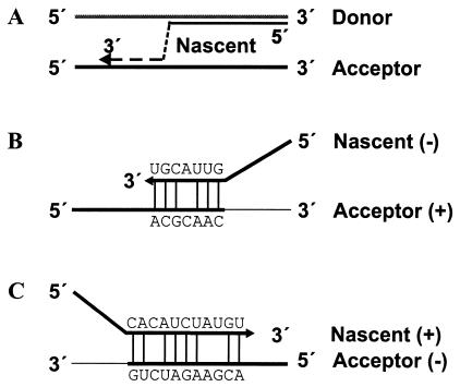
Four bovine viral diarrhea virus type 2 (BVDV-2) pairs consisting of cytopathogenic (cp) and noncp BVDV-2 were isolated during an outbreak of mucosal disease. Comparative sequence analysis showed that the four noncp BVDV-2 isolates were almost identical. For the cp BVDV-2 isolates, viral subgenomic RNAs were shown by Northern blot to have a length of about 8 kb, which is about 4.3 kb shorter than the genome of noncp BVDV. Cytopathogenicity and the expression of NS3 were both strictly correlated to the presence of viral subgenomic RNAs. By reverse transcription-PCR, Southern blot analysis, and nucleotide sequencing, a set of 11 unique subgenomes was identified with up to 5 different subgenomes isolated from one animal. To our knowledge, this is the first report on isolation of a set of pestiviral subgenomes from individual animals. Common features of the BVDV-2 subgenomic RNAs include (i) deletion of most of the genomic region encoding the structural proteins, as well as the nonstructural proteins p7 and NS2, and (ii) insertion of cellular (poly)ubiquitin coding sequences. Three subgenomes also comprised 15 to 75 nucleotides derived from the 5' part of the NS2 gene. Comparisons of the obtained nucleotide sequences revealed that the different BVDV-2 subgenomes evolved from the respective noncp BVDV-2 by RNA recombination. The presence of short regions of sequence similarity at several crossing-over sites suggests that base pairing between the nascent RNA strand and the acceptor RNA template facilitates template switching of the BVDV RNA-dependent RNA polymerase.


Figures

FIG. 1
FIG. 1
Expression of nonstructural proteins NS2-3 and NS3 (A) and detection of viral RNAs (B). (A) Immunoblot analysis. MDBK cells were infected with BVDV-2 Giessen-1 NCP-A, CP-A, CP-B, CP-C, or CP-D. The cells were lysed 48 h postinfection, and the samples were subjected to SDS-PAGE (8% polyacrylamide) under reducing conditions, transferred to a nitrocellulose membrane, and analyzed by using anti-NS3 monoclonal antibody 8.12.7. The sizes (in kilodaltons) of marker proteins are indicated on the left. The positions of NS2-3 and NS3 are marked with arrows. n.i., noninfected MDBK cells. (B) Northern blot analysis of total RNA from infected and noninfected MDBK cells. Prior to transfer and hybridization, RNA was separated on a 1.0% agarose gel under denaturing conditions. The blot was hybridized with a 2.5-kb NotI-NsiI fragment from the cDNA clone pA/BVDV (24). RNA ladder sizes in kilobases are indicated. Migration positions of the viral genomic and subgenomic RNAs are marked with arrows. The additional band with a size of ca. 4.5 kb is presumably due to the presence of large amounts of rRNA.
FIG. 2
FIG. 2
(A) RT-PCR strategy for identification of subgenomic viral RNAs with large deletions in the genomic region encoding Npro, the structural proteins, p7, and NS2. The positions and orientations of primers Ol P100 (▸) and Ol NS3R (◂) are indicated. The bull’s-eye icon indicates a putative insertion. NCP, noncp pestivirus. (B and C) RT-PCR products obtained from RNA of cells infected with BVDV-2 CP-A (lane 1), CP-B (lane 2), CP-C (lane 3), CP-D (lane 4), and NCP-A (lane 5). RNA from noninfected cells (−) served as a negative control. (B) The cDNA fragments were separated on a 1.0% agarose gel and stained with ethidium bromide. (C) Southern blot analysis of the agarose gel shown in panel B. After transfer to a nylon membrane, the blot was hybridized with a 2.5-kb NotI-NsiI fragment from the cDNA clone pA/BVDV (24). M, size standard; KB, kilobases.
FIG. 3
FIG. 3
Schematic representation of the genome organization of a noncp pestivirus (NCP) and the BVDV-2 subgenomic RNAs (only part of the genome/subgenomes is shown). The subgenomes A and B were isolated from animals A and B, respectively. C1, C2, C3, C4, and C5 were derived from animal C, while D1, D2, D3, and D4 were obtained from animal D. All subgenomic RNAs contain either the entire Npro gene or part of it (checkered box), as well as an insertion of cellular (poly)ubiquitin coding sequences (hatched box). The presence of capsid protein coding sequences (A, C3, C4, C5, and D4) is indicated by gray boxes. The presence of NS2 coding sequences (C2, D3, and D4) is indicated by black boxes. The numbers of amino acids corresponding to the respective viral protein(s) or (poly)ubiquitin are shown below the bars. The positions of the 5′ recombination junctions (⧫), the 3′ recombination junctions (●), and the additional internal recombination junctions of subgenomes C2, D3, and D4 (■) are indicated above the bars.
FIG. 4
FIG. 4
Alignment of sequences in the regions of the recombination junctions of BVDV-2 subgenomes. Nucleotide sequences of the parental template RNA molecules (upper and lower lines) are shown together with the sequences of the progeny subgenomic RNAs (middle lines). The sequence identity between the subgenomic RNAs and the respective template RNAs is indicated by the vertical lines between the sequences. The sites of recombination are indicated by either boxes (5′ sites of subgenomes C1, C3, D2, and D4 and the conserved 3′ site) or a vertical line (subgenome C4). The regions of sequence similarity between the nascent and the assumed acceptor RNA strand are highlighted by a gray background. The 3′ recombination site is conserved among all BVDV-2 subgenomes analyzed here. Recombination sites without regions of sequence similarity are not included.
FIG. 5
FIG. 5
(A) Replicase-mediated template switching model of RNA recombination. The model includes dissociation of the nascent RNA strand (3′ part as dashed line) from the donor template (gray line) followed by reassociation to an acceptor template (black line). (B and C) Sequence similarity-guided reassociation of the nascent RNA to the acceptor template during the synthesis of either negative (B)- or positive (C)-sense RNA strands. As examples, the sequences of the 5′ recombination junctions of subgenomes D4 (B) and D2 (C) and the reassociation regions of the respective acceptor templates are shown. Sequence complementarity between the nascent RNA and the acceptor template is indicated by the vertical lines between the sequences.

References

    1. Agol V I. Recombination and other genomic rearrangements in picornaviruses. Semin Virol. 1997;8:77–84.
    1. Becher P, König M, Paton D, Thiel H-J. Further characterization of border disease virus isolates: evidence for the presence of more than three species within the genus pestivirus. Virology. 1995;209:200–206. - PubMed
    1. Becher P, Meyers G, Shannon A D, Thiel H-J. Cytopathogenicity of border disease virus is correlated with integration of cellular sequences into the viral genome. J Virol. 1996;70:2992–2998. - PMC - PubMed
    1. Becher P, Orlich M, Shannon A D, Horner G, König M, Thiel H-J. Phylogenetic analysis of pestiviruses from domestic and wild ruminants. J Gen Virol. 1997;78:1357–1366. - PubMed
    1. Becher P, Orlich M, Thiel H-J. Complete genomic sequence of border disease virus, a pestivirus from sheep. J Virol. 1998;72:5165–5173. - PMC - PubMed

Publication types

MeSH terms

LinkOut - more resources