. 1999 Jul;73(7):5688-97.
doi: 10.1128/JVI.73.7.5688-5697.1999.

Epstein-Barr virus nuclear antigen 3C interacts with histone deacetylase to repress transcription

Affiliations

Epstein-Barr virus nuclear antigen 3C interacts with histone deacetylase to repress transcription

S A Radkov et al. J Virol. 1999 Jul.

Abstract

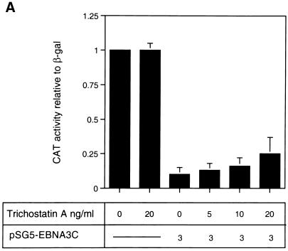
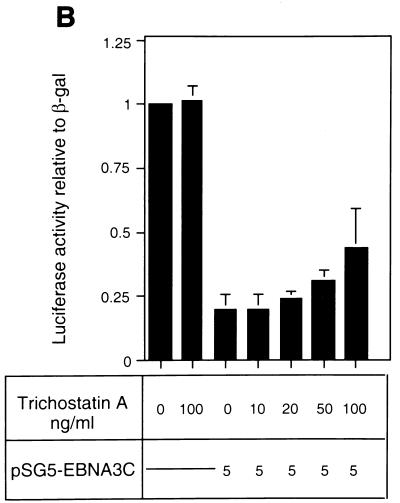
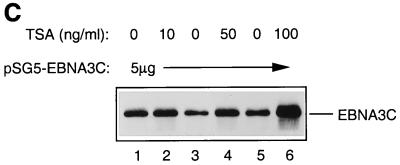
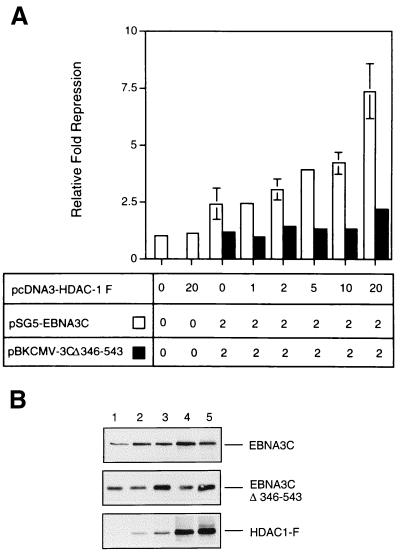
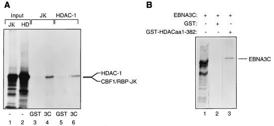
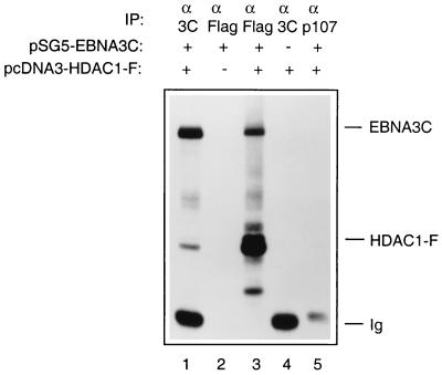
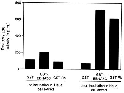
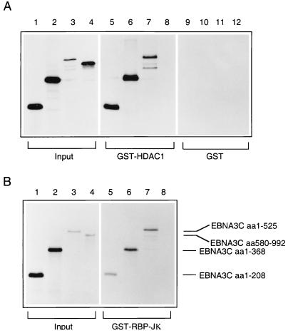
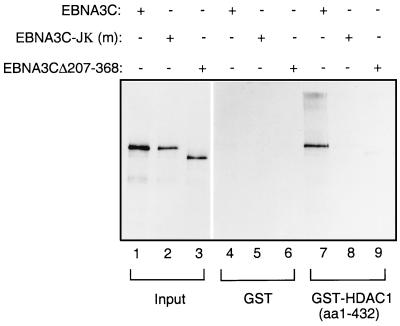
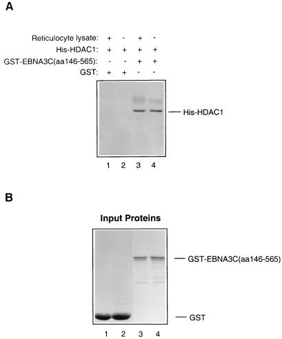
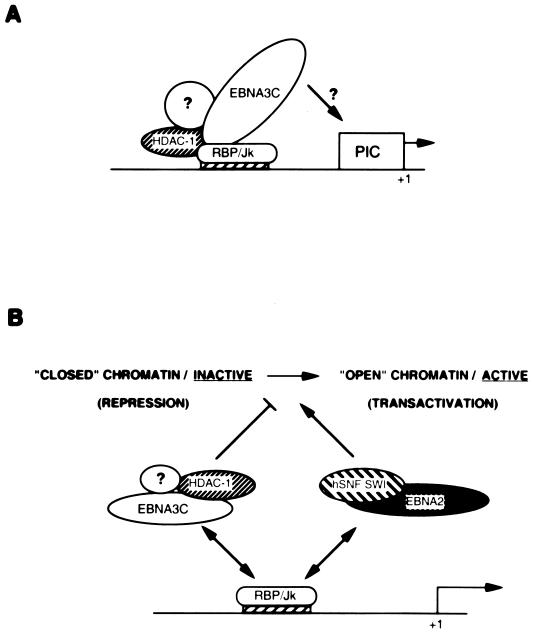
EBNA3C can specifically repress the expression of reporter plasmids containing EBV Cp latency-associated promoter elements. Cp is normally the main promoter for EBNA mRNA initiation, so it appears that EBNA3C contributes to a negative autoregulatory control loop. By mutational analysis it was previously established that this repression is consistent with EBNA3C being targeted to Cp by binding the cellular sequence-specific DNA-binding protein CBF1 (also known as recombination signal-binding protein [RBP]-Jkappa. Further analysis suggested that in vivo a corepressor interacts with EBNA3C in this DNA binding complex. Results presented here are all consistent with a component of such a corepressor exhibiting histone deacetylase activity. The drug trichostatin A, which specifically inhibits histone deacetylases, relieved two- to threefold the repression of Cp induced by EBNA3C in two different cell types. Moreover, repression of pTK-CAT-Cp4x by EBNA3C was specifically enhanced by cotransfection of an expression plasmid for human histone deacetylase-1 (HDAC1). Consistent with these functional assays, in vitro-translated HDAC1 bound to a glutathione S-transferase (GST) fusion protein including full-length EBNA3C, and in the reciprocal experiment EBNA3C bound to a GST fusion with the N terminus of HDAC1. Coimmunoprecipitations also revealed an EBNA3C-HDAC1 interaction in vivo, and GST-EBNA3C bound functional histone deacetylase enzyme activity from HeLa cell nuclear extracts. The region of EBNA3C involved in the interaction with HDAC1 appears to correspond to the region which is necessary for binding to CBF1/RBP-Jkappa. A direct physical interaction between EBNA3C and HDAC1 was demonstrated with recombinant proteins purified from bacterial cells, and we therefore conclude that HDAC1 and CBF1/RBP-Jkappa bind to the same or adjacent regions of EBNA3C. These data suggest that recruitment of histone deacetylase activity makes a significant contribution to the repression of transcription from Cp because EBNA3C bridges an interaction between CBF1/RBP-Jkappa and HDAC1.


Figures

FIG. 1
FIG. 1
(A) Effect of TSA on EBNA3C-mediated repression in DG75 B cells. DG75 cells were transfected with 2 μg of the pTK-CAT-Cp4× reporter plasmid and with the pSG5-EBNA3C effector plasmid as indicated. Transfected cells were treated for 24 h with the amounts of TSA shown. CAT assays were performed and standardized to β-galactosidase activity expressed from the cotransfected plasmid pSV-β-Gal (2 μg). The data shown are relative to the activity from untreated pTK-CAT-Cp4×, which was given an arbitrary value of 1. Each result is the mean and standard deviation from three independent experiments. (B) Effect of TSA on EBNA3C-mediated repression in Jurkat T cells. Jurkat cells were transfected with 2 μg of the p1425-Cp-Luc reporter plasmid and with the pSG5-EBNA3C effector plasmid as indicated. Transfected cells were treated with the indicated amounts of TSA for 24 h. Luciferase assays were performed and standardized to β-galactosidase activity expressed from the cotransfected plasmid pSV-β-Gal (2 μg). Data shown are relative to the activity from untreated p1425-Cp-Luc, which was given an arbitrary value of 1. Each result is the mean and standard deviation from three independent experiments. (C) Western blot analysis of EBNA3C in a representative experiment performed with p1425-Cp-Luc in Jurkat cells. The protein extract from transfected cells was resolved by SDS-PAGE (7.5% polyacrylamide). After transfer to a nitrocellulose membrane, the proteins were probed with the anti-EBNA3C MAb A10.
FIG. 1
FIG. 1
(A) Effect of TSA on EBNA3C-mediated repression in DG75 B cells. DG75 cells were transfected with 2 μg of the pTK-CAT-Cp4× reporter plasmid and with the pSG5-EBNA3C effector plasmid as indicated. Transfected cells were treated for 24 h with the amounts of TSA shown. CAT assays were performed and standardized to β-galactosidase activity expressed from the cotransfected plasmid pSV-β-Gal (2 μg). The data shown are relative to the activity from untreated pTK-CAT-Cp4×, which was given an arbitrary value of 1. Each result is the mean and standard deviation from three independent experiments. (B) Effect of TSA on EBNA3C-mediated repression in Jurkat T cells. Jurkat cells were transfected with 2 μg of the p1425-Cp-Luc reporter plasmid and with the pSG5-EBNA3C effector plasmid as indicated. Transfected cells were treated with the indicated amounts of TSA for 24 h. Luciferase assays were performed and standardized to β-galactosidase activity expressed from the cotransfected plasmid pSV-β-Gal (2 μg). Data shown are relative to the activity from untreated p1425-Cp-Luc, which was given an arbitrary value of 1. Each result is the mean and standard deviation from three independent experiments. (C) Western blot analysis of EBNA3C in a representative experiment performed with p1425-Cp-Luc in Jurkat cells. The protein extract from transfected cells was resolved by SDS-PAGE (7.5% polyacrylamide). After transfer to a nitrocellulose membrane, the proteins were probed with the anti-EBNA3C MAb A10.
FIG. 2
FIG. 2
(A) Repression by EBNA3C is enhanced by HDAC1 expression. DG75 cells were transiently transfected with 2 μg of the pTK-CAT-Cp4× reporter plasmid, 2 μg of pSV-β-Gal, and 2 μg of either the pSG5-EBNA3C or the pBKCMV-EBNA3CΔ346-543 effector plasmid. The indicated amount (in micrograms) of pcDNA3–HDAC-1F was added to each transfection. Cell extracts were prepared 48 h after transfection, and CAT activity was determined. After normalization to β-galactosidase activity, the data were expressed as fold repression relative to the activity from pTK-CAT-Cp4× with empty control vectors, which was given an arbitrary value of 1. Means and standard deviations from three independent experiments are shown. (B) Western blot analysis of the expression of EBNA3C, EBNA3CΔ346-543, and flag-tagged HDAC1 in a representative experiment. Protein extract was resolved by SDS-PAGE (10% polyacrylamide). After transfer to a nitrocellulose membrane, the proteins were probed with the anti-EBNA3C MAb A10 and the anti-flag antibody D-8. Lane 1, 0 μg of pcDNA3–HDAC1-F DNA; lanes 2 through 5, 1, 2, 10, and 20 μg, respectively.
FIG. 3
FIG. 3
(A) HDAC1 interacts with GST-EBNA3C. GST-EBNA3C expressed from a baculovirus was incubated with equal amounts of [35S]methionine-labelled RBP-Jκ (as a positive control) (lane 4) or HDAC1 (lane 6). Bound proteins were resolved by SDS-PAGE (7.5% polyacrylamide gel). Lanes 1 and 2 contain 10% of the input protein in each binding reaction, and lanes 3 and 5 contain the protein which bound to GST. (B) EBNA3C interacts with the N terminus of HDAC1 fused to GST. Bacterially expressed GST-HDAC1 (aa 1 to 382) was incubated with equal amounts of [35S]methionine-labelled EBNA3C (lane 3). Bound proteins were resolved by SDS-PAGE (7.5% polyacrylamide gel). Lane 1 contains 10% of the input protein in each binding reaction, and lane 2 contains the protein which bound to GST.
FIG. 4
FIG. 4
EBNA3C and HDAC1 can interact in vivo. DG75 cells were transfected with a mixture of 5 μg of pSG5-EBNA3C and 5 μg of pcDNA–HDAC1-F (lanes 1, 3, and 5) or with pSG5-EBNA3C alone (lane 2) or pcDNA3–HDAC1-F alone (lane 4). Cells were harvested 48 h after transfection and immunoprecipitated (IP) with the appropriate MAbs as indicated. After separation on an SDS–7.5% polyacrylamide gel, the proteins were Western blotted onto a nitrocellulose membrane and probed sequentially with anti-EBNA3C (MAb A10) and anti-flag (MAb D-8).
FIG. 5
FIG. 5
EBNA3C can precipitate histone deacetylase enzyme activity from HeLa nuclear extracts. Nuclear extracts from HeLa cells were subjected to a GST pulldown analysis with beads coated with GST-EBNA3C, GST-Rb, or wild-type GST alone. In a parallel control experiment, the coated beads were incubated without nuclear extract in the reaction mixtures. Bound proteins were assayed for histone deacetylase activity as described in Materials and Methods. All samples were assayed in duplicate.
FIG. 6
FIG. 6
Both HDAC1 and RBP-Jκ interact with the N terminus of EBNA3C. (A) In a GST pulldown experiment, bacterially expressed GST-HDAC1 (aa 1 to 432) was incubated with equal amounts of [35S]methionine-labelled EBNA3C deletion mutants: aa 1 to 208 (lane 5), aa 1 to 368 (lane 6), aa 1 to 525 (lane 7), and aa 580 to 992 (lane 8). The protein pulled down by wild-type GST is shown in lanes 9 through 12. (B) In a parallel pulldown experiment, the same EBNA3C deletion mutants were incubated with bacterially expressed GST-RBP-Jκ. In both experiments, bound proteins were resolved by SDS-PAGE (7.5% polyacrylamide), and comparable amounts of GST-HDAC1 and GST-RBP-Jκ were used in the binding reactions (data not shown). In both experiments, lanes 1 through 4 contain 10% of the input protein included in each binding reaction.
FIG. 7
FIG. 7
HDAC1 binds to the region of EBNA3C which interacts with CBF1/RBP-Jκ. In a GST pulldown experiment, bacterially expressed GST-HDAC1 (aa 1 to 432) was incubated with equal amounts of [35S]methionine-labelled EBNA3C (lane 7); EBNA3C-Jκ (m), a mutant which no longer binds CBF1/RBP-Jκ (lane 8); and EBNA3CΔ207-368, which also fails to bind CBF1/RBP-Jκ (lane 9). Lanes 4 through 6 contain protein bound by wild-type GST, and lanes 1 through 3 show 10% of the input protein in each binding reaction.
FIG. 8
FIG. 8
EBNA3C binds directly to HDAC1. (A) Equal amounts of His-HDAC1, purified from a bacterial lysate, were incubated with Sepharose beads loaded with GST-EBNA3C (aa 146 to 565) or GST. Similar binding reactions were set up with a supplement of TNT reticulocyte lysate (Promega). After washing, the bound proteins were resolved by SDS-PAGE (12% polyacrylamide), transferred to a nitrocellulose membrane, and probed with a polyclonal antiserum which recognizes the C terminus of HDAC1. His-HDAC1 bound to GST-EBNA3C (aa 146 to 565) (lanes 3 and 4) irrespective of whether reticulocyte lysate was present. No binding to GST was detected (lanes 1 and 2). (B) Brilliant blue-stained polyacrylamide gel showing the amounts of purified protein used in each binding reaction.
FIG. 9
FIG. 9
A possible role for EBNA3C in modulating transcription. (A) All the data reported previously (21, 46) and in this study are consistent with a model in which CBF1/RBP-Jκ can reversibly associate with HDAC1 and target it to DNA. Since EBNA3C binds to both proteins, we propose that EBNA3C can bridge this interaction and so mediate repression. It is unclear whether the other members of the deacetylase repression machinery (such as SMRT or Sin3) are also associated with HDAC1 in these complexes or whether EBNA3C establishes a novel complex with deacetylase activity. It is also unclear whether EBNA3C additionally contributes to repression by interacting with the basal transcription machinery (represented by the preinitiation complex [PIC]). (B) In cells latently infected with EBV, there will be a dynamic equilibrium between EBNA3C–HDAC1–RBP-Jκ complexes and EBNA2–hSNF-SWI–RBP-Jκ complexes (50) associated with Cp, so the surrounding chromatin configuration will be in a state of flux. The net result will be finely regulated EBNA expression. It should be noted that EBNA3A and EBNA3B (which can also bind RBP-Jκ) may also influence the protein-protein and protein-DNA interactions on Cp. For the sake of simplicity, the other factors which associate with EBNA2 have not been included, and neither have the endogenous cell factors TAN-1 and activated Notch receptor, which may also influence chromatin conformation in a manner analogous to that of EBNA2 (21).

Similar articles

Cited by

References

    1. Alfieri C, Birckenbach M, Kieff E. Early events in Epstein-Barr virus infection of human lymphocytes. Virology. 1991;181:595–608. - PubMed
    1. Alland L, Muhle R, Hou H, Potes J, Chin L, Schreiber-Agus N, DePinho R. Role for N-CoR and histone deacetylase in Sin3-mediated transcriptional repression. Nature. 1997;387:49–55. - PubMed
    1. Allday M J, Crawford D H, Griffin B E. Prediction and demonstration of a novel Epstein-Barr virus nuclear antigen. Nucleic Acids Res. 1988;16:4353–4367. - PMC - PubMed
    1. Allday M J, Crawford D H, Griffin B E. Epstein-Barr virus latent gene expression during the initiation of B cell immortalisation. J Gen Virol. 1989;70:1755–1764. - PubMed
    1. Ayer D E, Lawrence Q A, Eisenman R N. Mad-Max transcriptional repression is mediated by ternary complex formation with mammalian homologs of yeast repressor Sin3. Cell. 1995;80:767–776. - PubMed

Publication types

MeSH terms