. 1999 Jul;73(7):5814-25.
doi: 10.1128/JVI.73.7.5814-5825.1999.

Induction of AIDS in rhesus monkeys by a recombinant simian immunodeficiency virus expressing nef of human immunodeficiency virus type 1

Affiliations

Induction of AIDS in rhesus monkeys by a recombinant simian immunodeficiency virus expressing nef of human immunodeficiency virus type 1

L Alexander et al. J Virol. 1999 Jul.

Abstract

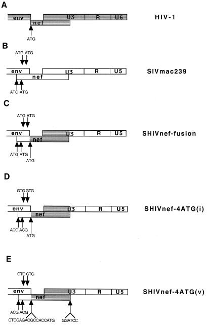
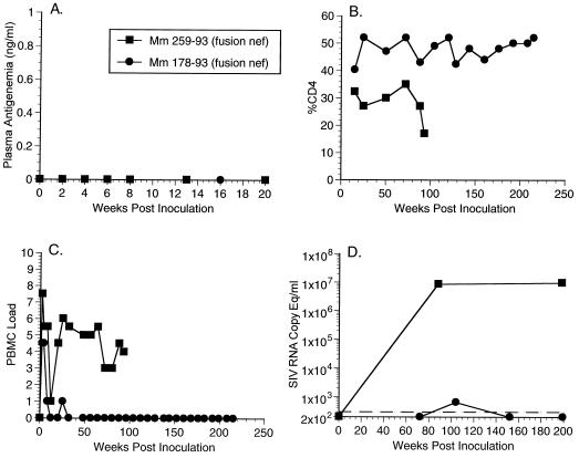
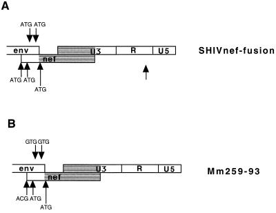
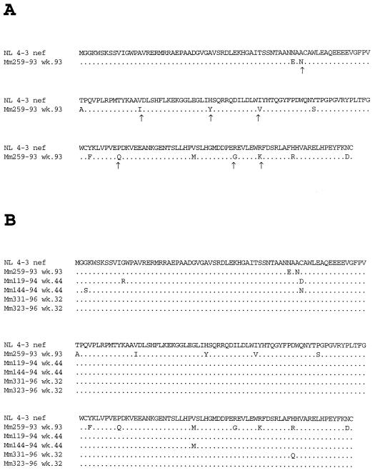
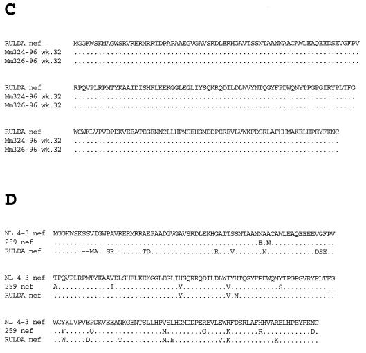
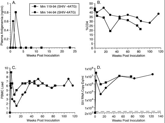
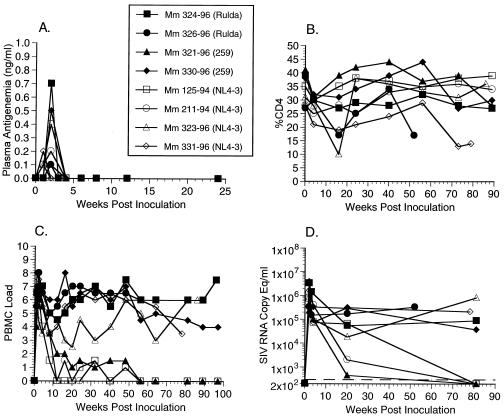
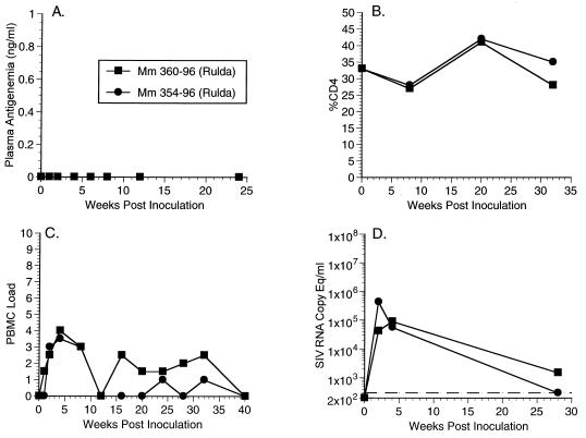
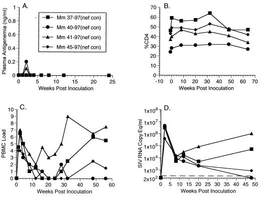
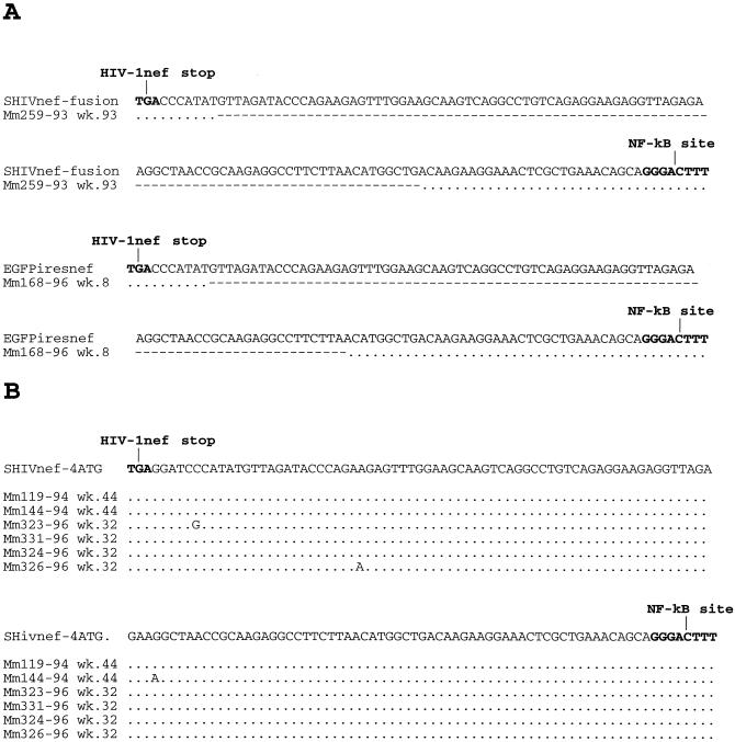
A nef gene is present in all primate lentiviruses, including human immunodeficiency virus type 1 (HIV-1) and simian immunodeficiency virus of macaque monkeys (SIVmac). However, the nef genes of HIV-1 and SIVmac exhibit minimal sequence identity, and not all properties are shared by the two. Nef sequences of SIVmac239 were replaced by four independent nef alleles of HIV-1 in a context that was optimal for expression. The sources of the HIV-1 nef sequences included NL 4-3, a variant NL 4-3 gene derived from a recombinant-infected rhesus monkey, a patient nef allele, and a nef consensus sequence. Of 16 rhesus monkeys infected with these SHIVnef chimeras, 9 maintained high viral loads for prolonged periods, as observed with the parental SIVmac239, and 6 have died with AIDS 52 to 110 weeks postinfection. Persistent high loads were observed at similar frequencies with the four different SIV recombinants that expressed these independent HIV-1 nef alleles. Infection with other recombinant SHIVnef constructions resulted in sequence changes in infected monkeys that either created an open nef reading frame or optimized the HIV-1 nef translational context. The HIV-1 nef gene was uniformly retained in all SHIVnef-infected monkeys. These results demonstrate that HIV-1 nef can substitute for SIVmac nef in vivo to produce a pathogenic infection. However, the model suffers from an inability to consistently obtain persisting high viral loads in 100% of the infected animals, as is observed with the parental SIVmac239.


Figures

FIG. 1
FIG. 1
Diagrams of HIV-1, SIVmac, and SHIVnef genomes. The gray areas represent HIV-1 sequences, and the white areas represent SIVmac sequences. The arrows indicate the sequence contained in different SHIVnef recombinants. The arrows below the nef gene indicate ATGs contained in the nef reading frame, and the arrows above the nef gene indicate ATGs contained in the non-env/non-nef reading frame in the region of overlap containing both SIV env and SIV nef. The genes or genetic elements depicted in this figure are not drawn to scale.
FIG. 2
FIG. 2
Plasma antigenemia, CD4%, PBMC load, and RNA copy eq/ml measurements for animal experiment 542 (AE542, Table 1). (A) Plasma antigenemia in SHIVnef-infected rhesus monkeys. p27 concentrations in plasma were determined at the time points indicated. The limit of detection is approximately 0.05 ng/ml. Week 0 is a preinfection sample taken immediately before inoculation with SHIVnef. (B) CD4% in SHIVnef-infected rhesus monkeys. Whole blood was drawn from SHIVnef-inoculated animals at various times p.i. and stained with OKT4, an FITC-conjugated murine monoclonal antibody that was raised against rhesus macaque CD4 (American Type Culture Collection). The stained samples were analyzed with a FACSscan flow cytometer (Becton Dickinson). (C) Frequency of infectious cells in PBMC of SHIVnef-infected rhesus macaques. Viral loads were graded on a scale from 0 to 10 indicating the number of PBMC needed to recover SIV. A “0” indicates that no virus was recovered with 106 cells, a “1” indicates successful virus recovery from 106 cells, and values 2 to 10 indicate successful virus recovery from 333,333, 111,111, 37,037, 12,345, 4,115, 1,371, 457, 152, or 51 cells, respectively. (D) Plasma SIV RNA levels at the indicated weeks p.i. for animals infected with SHIVnef recombinants. The dashed lines indicate the threshold sensitivity of the assay (300 copy eq/ml). Prior to week 72, plasma was not stored appropriately for plasma RNA measurements of animal experiment 542 (AE542). A value of 0 was assumed for week 0.
FIG. 3
FIG. 3
Sequence changes in the region of overlap containing both SIV env and SIV nef. SHIVnef sequences present in PBMC of Mm259-93 at the time of death are compared with the SHIVnef fusion construct that was used for infection. One of the preserved ATGs represents the initiating ATG of HIV-1 nef. The other preserved ATG in the region of overlap is not in an appropriate consensus (5′-TCCATGA-3′) context for the initiation of translation (a purine at the −3 position and a guanosine at the +4 position relative to the adenosine of the ATG) (–25). These data represent a consensus sequence of two clones from each of two independent PCRs.
FIG. 4
FIG. 4
Analysis of HIV-1 nef sequences contained in SHIVnef recombinants recovered from infected monkeys. The sequences represent a consensus of two clones from each of two independent PCRs. (A) Amino acid changes observed in NL 4-3 nef after infection of Mm259-93 with SHIVnef-fusion. The arrows indicate where nucleotide changes resulted in the same amino acid that is present at the corresponding position of SIVmac nef. (B) A comparison of HIV-1 nef amino acid sequences after passage in animals that were infected with SHIVnef that expressed NL 4-3 nef sequences and maintained high viral loads. (C) Same as in panel B except with RULDA nef sequences. (D) An alignment of the amino acids expressed by NL 4-3 nef, 259 nef which was passaged through Mm259-93, and RULDA nef which was isolated from a rapid-progressor HIV-1 patient. In all panels, dots denote sequence identity and the dashes denote deletions. The sequences analyzed were isolated from these animals at the indicated number of weeks p.i.
FIG. 4
FIG. 4
Analysis of HIV-1 nef sequences contained in SHIVnef recombinants recovered from infected monkeys. The sequences represent a consensus of two clones from each of two independent PCRs. (A) Amino acid changes observed in NL 4-3 nef after infection of Mm259-93 with SHIVnef-fusion. The arrows indicate where nucleotide changes resulted in the same amino acid that is present at the corresponding position of SIVmac nef. (B) A comparison of HIV-1 nef amino acid sequences after passage in animals that were infected with SHIVnef that expressed NL 4-3 nef sequences and maintained high viral loads. (C) Same as in panel B except with RULDA nef sequences. (D) An alignment of the amino acids expressed by NL 4-3 nef, 259 nef which was passaged through Mm259-93, and RULDA nef which was isolated from a rapid-progressor HIV-1 patient. In all panels, dots denote sequence identity and the dashes denote deletions. The sequences analyzed were isolated from these animals at the indicated number of weeks p.i.
FIG. 5
FIG. 5
Plasma antigenemia, CD4%, PBMC load, and RNA copy eq/ml measurements for animal experiment 576 (AE576, Table 1). See legend to Fig. 2 for details.
FIG. 6
FIG. 6
Plasma antigenemia, CD4%, PBMC load, and RNA copy eq/ml measurements for animal experiment 585 (AE585, Table 1). See legend to Fig. 2 for details.
FIG. 7
FIG. 7
Plasma antigenemia, CD4%, PBMC load, and RNA copy eq/ml measurements for animal experiment 603 (AE603, Table 1). See legend to Fig. 2 for details.
FIG. 8
FIG. 8
Plasma antigenemia, CD4%, PBMC load, and RNA copy eq/ml measurements for animal experiment 598 (AE598, Table 1). See legend to Fig. 2 for details.
FIG. 9
FIG. 9
Infection of CEMx174 cells with SIVmac239 and various SHIVnef recombinants. A 1-ng portion of p27 antigen that was derived from the cell-free supernatant of transfected CEMx174 cells was used for each infection. Depicted are the SIV p27 concentrations in the cell-free supernatants of the infected cells. Symbols: ■, SIVmac239; ●, SHIVnef NL 4-3v; ▴, SHIVnef RULDA; ⧫, SHIVnef259; □, SHIVnef consensus; ○, SHIVnef fusion.
FIG. 10
FIG. 10
Infection of unstimulated 221 cells with SHIVnef recombinants. Cell line 221 cells incubated in RPMI medium containing 5% fetal calf serum without exogenous IL-2 were infected with 10 ng of SIV p27 antigen of SHIVnef recombinant virus. p27 antigen was quantitated in the cell-free supernatant at the times indicated. Symbols: ■, SIVmac239; ●, SHIVnef-fusion; ▴, SHIVnef-4ATG(i); ⧫, SHIVnef NL 4-3v; □, SIVmac239Δnef; ○, SHIVnef(i); ▵, SHIVnef-2ATG.
FIG. 11
FIG. 11
Expression of HIV-1 nef in CEMx174 cells infected with SHIVnef recombinants. HIV-1- or SHIVnef-infected cells were harvested at day 7, and proteins from cell lysates were separated by SDS-PAGE and electroblotted onto a membrane filter. HIV-1 nef was detected by using a nef-specific monoclonal antiserum (EH). Lanes: 1, mock infected; 2, HIV-1 NL 4-3 nef; 3, SHIVnef(i); 4, SHIVnef-stop; 5, SHIVnef-fusion; 6, SHIVnef-2Met; 7, SHIVnef-4ATG(i); 8, SHIVnef-4ATG(v).
FIG. 12
FIG. 12
Sequence analysis of SHIVnef U3 sequences before and after passage in rhesus monkeys. Dots denote no change in the U3 sequence from the inoculum, and dashes denote deletions in the U3 sequence after animal infection.

References

    1. Adachi A, Gendelman H E, Koenig S, Folks T, Willey R, Rabson A, Martin M. Production of acquired immunodeficiency syndrome-associated retrovirus in human and nonhuman cells transfected with an infectious molecular clone. J Virol. 1986;59:284–291. - PMC - PubMed
    1. Aiken C, Konner J, Landau N, Lenburg M E, Trono D. Nef induces CD4 endocytosis: requirement for a critical dileucine motif in the membrane-proximal CD4 cytoplasmic domain. Cell. 1994;76:853–864. - PubMed
    1. Aiken C, Trono D. Nef stimulates human immunodeficiency virus type 1 proviral DNA synthesis. J Virol. 1995;69:5048–5056. - PMC - PubMed
    1. Alexander L, Du Z, Rosenzweig M, Jung J U, Desrosiers R C. A role for natural simian immunodeficiency virus and human immunodeficiency virus type 1 nef alleles in lymphocyte activation. J Virol. 1997;71:6094–6099. - PMC - PubMed
    1. Alexander L, Veazey R S, Czajak S, DeMaria M, Rosenzweig M, Lackner A A, Desrosiers R C, Sasseville V G. Recombinant simian immunodeficiency virus expressing green fluorescent protein identifies infected cells in rhesus monkeys. AIDS Res Hum Retroviruses. 1999;15:11–21. - PubMed

Publication types

Substances