. 1999 Jul;73(7):5852-64.
doi: 10.1128/JVI.73.7.5852-5864.1999.

Role of the M2-1 transcription antitermination protein of respiratory syncytial virus in sequential transcription

Affiliations

Role of the M2-1 transcription antitermination protein of respiratory syncytial virus in sequential transcription

R Fearns et al. J Virol. 1999 Jul.

Abstract

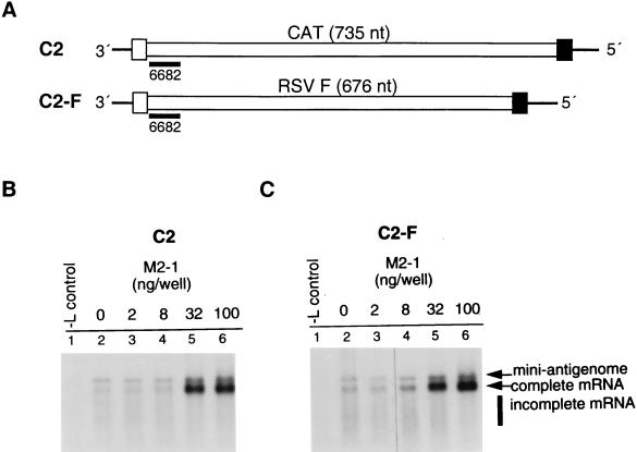
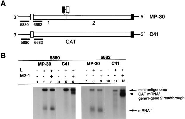
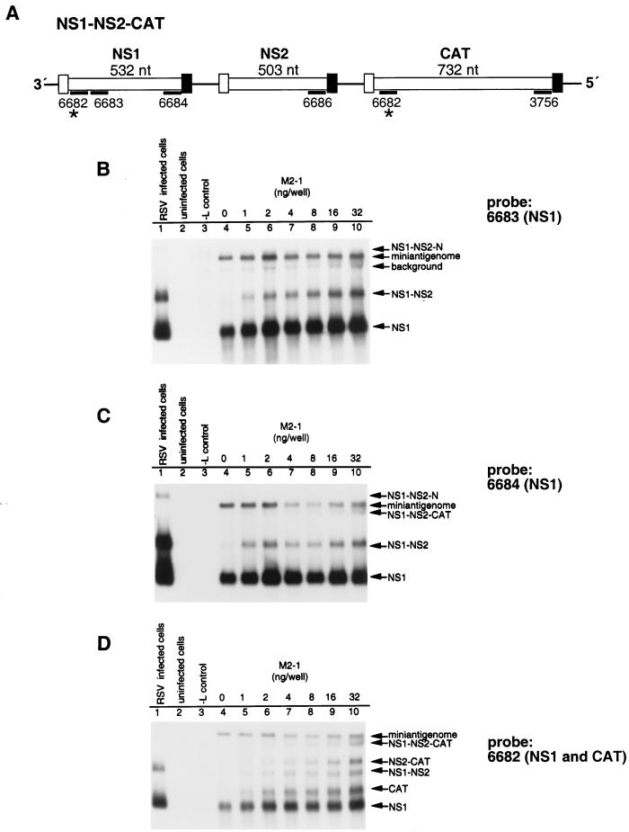
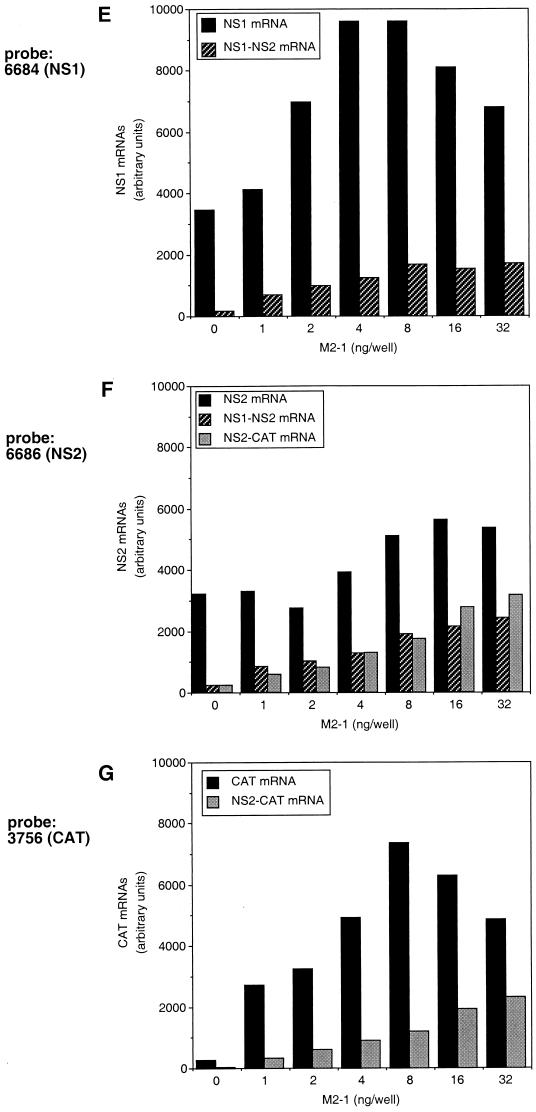
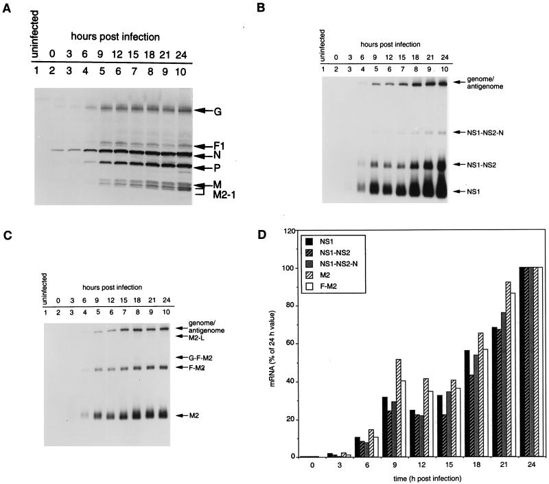
M2-1 protein of human respiratory syncytial virus (RSV) is a transcription antitermination factor that is important for the efficient synthesis of full-length mRNAs as well as for the synthesis of polycistronic readthrough mRNAs, which are characteristic of nonsegmented negative-strand RNA viruses. The contributions of these effects to RSV sequential transcription were investigated with minigenomes which contained one to five genes which were either foreign marker genes or authentic RSV genes. When evaluated on a promoter-proximal gene, the effect of M2-1 on the synthesis of full-length mRNA was much greater for a long (1,212- or 1,780-nucleotide) gene (up to a 615-fold increase) than for a short (274-nucleotide) gene (less than a 2-fold increase). This was independent of whether the gene contained non-RSV or RSV-specific sequence. Once the polymerase had terminated prematurely, it was unable to reinitiate at a downstream gene. These studies also confirmed that M2-1 enhances the synthesis of polycistronic mRNAs and that the magnitude of this effect varied greatly among different naturally occurring gene junctions. The synthesis of polycistronic mRNAs, which presumably involves antitermination at the gene-end signal, required a higher level of M2-1 than did the synthesis of the corresponding monocistronic mRNAs. M2-1 did not have a comparable antitermination effect at the junction between the leader region and the first gene. In a minigenome containing the NS1 and NS2 genes in their authentic sequence context, synthesis of full-length NS1 and NS2 mRNAs in the absence of M2-1 was remarkably high (36 and 57%, respectively, of the maximum levels observed in the presence of M2-1). In contrast, synthesis of mRNA from additional downstream genes was highly dependent on M2-1. Thus, RSV has the potential for two transcription programs: one in the absence of M2-1, in which only the NS1 and NS2 genes are transcribed, and one in the presence of M2-1, in which sequential transcription of the complete genome occurs. The dependence on M2-1 for transcription was greater for a gene in the fifth position from the promoter than for one in the third position. This indicates that under conditions where M2-1 is limiting, its concentration affects the gradient of transcription. Although M2-1 was found to have profound effects on transcription, it had no effect on replication of any minigenome tested, suggesting that it is not an active participant in RNA replication or regulation of RNA replication. Finally, since a permissive RSV infection is marked by a gradual increase in the intracellular accumulation of viral proteins including M2-1, we examined the relative abundances of various mRNAs during RSV infection for evidence of temporal regulation of transcription. None was found, implying that the availability of M2-1 during a permissive infection is sufficient at all times such that its concentration does not mediate temporal regulation of gene transcription.


Figures

FIG. 1
FIG. 1
In the absence of M2-1, the RSV polymerase efficiently synthesizes short, but not long, mRNAs and does not reinitiate following intragenic termination. (A) Structures of the MP-30 and RF-9 minigenomes. Minigenome MP-30 is a dicistronic minigenome in which the CAT coding sequence has been broken into two genes of 274 and 495 nt separated by the N-P gene junction of RSV (see Materials and Methods). Minigenome RF-9 was constructed by inserting the RSV F sequence (hatched) into the first gene of MP-30 (dotted lines). GS and GE transcription signals are shown as small open and solid boxes, respectively; negative-sense oligonucleotide probes 6682, 5629, 5878, and 3756 are shown as short thick lines. These conventions are used in each figure. (B and C) Northern blot analyses of positive-sense RNAs synthesized in HEp-2 cells which were infected with vaccinia virus recombinant vTF7-3 and simultaneously transfected with plasmid MP-30 (B) or RF-9 (C), and the support plasmids N and P, together with the indicated combinations of L and M2-1. RNA was purified 48 h later, subjected to electrophoresis on formaldehyde gels, transferred to nitrocellulose, and analyzed by hybridization with the indicated 5′-end-labelled oligonucleotide.
FIG. 2
FIG. 2
M2-1 is required for fully processive transcription of authentic RSV sequence as well as foreign sequence. (A) Structures of minigenomes C2, in which the gene is composed mainly of foreign CAT sequence, and C2-F, in which the gene is composed solely of RSV-specific sequence. The negative-sense oligonucleotide, 6682, hybridizes to the nontranslated region of NS1, which forms the 5′ end of each mRNA. (B and C) Northern blot analysis of positive-sense RNAs synthesized in HEp-2 cells which were transfected as described for Fig. 1 with plasmid encoding minigenome C2 (B) or C2-F (C) together with plasmids N and P (lane 1), N, P, and L (lane 2), or N, P, or L, and the indicated amounts of M2-1 (lanes 3 to 6). RNA was analyzed by Northern blot hybridization to oligonucleotide 6682.
FIG. 3
FIG. 3
Lack of effect of M2-1 on events at the leader gene junction. (A) Structures of minigenomes MP-30 and C41. Minigenome MP-30 is identical to C41 except that it contains the N-P gene junction inserted within the CAT gene. Oligonucleotide 5880 is specific against the positive-sense leader transcript, and oligonucleotide 6682 hybridizes to the nontranslated region of NS1. (B) Northern blot analysis of positive-sense RNAs from HEp-2 cells which were transfected as described for Fig. 1 with plasmid MP-30 or C41 and the support plasmids N and P, together with the indicated combinations of L and M2-1. RNA was analyzed by Northern blot hybridization with the indicated 5′-end-labelled oligonucleotide.
FIG. 4
FIG. 4
Expression of positive-sense RNAs from a tricistronic minigenome, NS1-NS2-CAT, which contains the 3′-terminal 1,125 nt of the RSV genome, including the NS1 and NS2 genes, followed by the CAT gene. (A) Diagram of minigenome NS1-NS2-CAT and locations of the negative-sense oligonucleotide probes. Oligonucleotide 6682 (∗) hybridizes to the nontranslated region of NS1, which is represented twice in the NS1-NS2-CAT minigenome and thus detects the NS1 and CAT mRNAs. (B to D) Northern blot analyses of positive-sense RNAs synthesized in HEp-2 cells transfected with NS1-NS2-CAT plasmid together with plasmids N and P (lane 3), N, P, and L (lane 4), or N, P, L, and the indicated amounts of M2-1 plasmid (lanes 5 to 10). Lane 1 contains total RNA isolated from RSV-infected HEp-2 cells at 24 h postinfection, and lane 2 contains RNA isolated from uninfected cells. Hybridization was performed with the indicated oligonucleotide probe. (E to G) Quantitation of the mRNAs detected with oligonucleotides 6684, 6686, and 3756, respectively, which each hybridize to the downstream end of one of the three mRNAs. The RNA bands in each lane were normalized so that the mini-antigenome equalled 1,000 units.
FIG. 4
FIG. 4
Expression of positive-sense RNAs from a tricistronic minigenome, NS1-NS2-CAT, which contains the 3′-terminal 1,125 nt of the RSV genome, including the NS1 and NS2 genes, followed by the CAT gene. (A) Diagram of minigenome NS1-NS2-CAT and locations of the negative-sense oligonucleotide probes. Oligonucleotide 6682 (∗) hybridizes to the nontranslated region of NS1, which is represented twice in the NS1-NS2-CAT minigenome and thus detects the NS1 and CAT mRNAs. (B to D) Northern blot analyses of positive-sense RNAs synthesized in HEp-2 cells transfected with NS1-NS2-CAT plasmid together with plasmids N and P (lane 3), N, P, and L (lane 4), or N, P, L, and the indicated amounts of M2-1 plasmid (lanes 5 to 10). Lane 1 contains total RNA isolated from RSV-infected HEp-2 cells at 24 h postinfection, and lane 2 contains RNA isolated from uninfected cells. Hybridization was performed with the indicated oligonucleotide probe. (E to G) Quantitation of the mRNAs detected with oligonucleotides 6684, 6686, and 3756, respectively, which each hybridize to the downstream end of one of the three mRNAs. The RNA bands in each lane were normalized so that the mini-antigenome equalled 1,000 units.
FIG. 5
FIG. 5
M2-1 alters the transcription gradient. (A) Structures of minigenomes NS1-NS2-N/CAT and NS1-NS2-N-P-M/CAT. The first two genes and gene junctions of NS1-NS2-N/CAT are identical to those of recombinant RSV, and the third gene is a chimera which contains 1,005 nt of the N gene of RSV fused to CAT. Likewise, NS1-NS2-N-P-M/CAT contains the first four genes and gene junctions of RSV, and the fifth gene consists of 180 nt of the RSV M gene fused to CAT. (B to E) Northern blots of positive-sense RNAs synthesized in HEp-2 cells transfected with plasmid encoding either minigenome NS1-NS2-N/CAT (B and C) or NS1-NS2-N-P-M/CAT (D and E) and the support plasmids N and P (lane 10), N, P, and L (lane 2), or N, P, L, and the indicated amounts of M2-1 plasmid (lanes 3 to 9). Lane 1 contains total RNA from RSV-infected cells (panels B and D) or from cells which received plasmids encoding minigenome C2, N, P, L, and 100 ng of M2-1 (panels C and E).
FIG. 6
FIG. 6
Lack of temporal regulation of RSV transcription. HEp-2 cells were infected with RSV (A2) at a multiplicity of infection of 4, and samples were harvested for RNA and protein at 3-h intervals. (A) Western blot of protein samples isolated from 0 to 24 h postinfection (lanes 2 to 10). Lane 1 contained total protein from uninfected cells. The RSV proteins were detected with an antiserum raised against gradient purified RSV virions. (B and C) Northern blots of RNA samples isolated simultaneously with the protein samples and arranged in the same lane order. The blots in panels B and C were hybridized with double-stranded DNA probes labelled by random priming and specific to NS1 and M2, respectively. (D) For each time point, the amount of each mRNA was normalized relative to genome-antigenome as an internal standard and expressed relative to the 24-h time point as 100.

References

    1. Abraham G, Banerjee A K. Sequential transcription of the genes of vesicular stomatitis virus. Proc Natl Acad Sci USA. 1976;73:1504–1508. - PMC - PubMed
    1. Atreya P L, Peeples M E, Collins P L. The NS1 protein of human respiratory syncytial virus is a potent inhibitor of minigenome transcription and RNA replication. J Virol. 1998;72:1452–1461. - PMC - PubMed
    1. Ball L A, White C N. Order of transcription of genes of vesicular stomatitis virus. Proc Natl Acad Sci USA. 1976;73:442–446. - PMC - PubMed
    1. Barik S. Transcription of human respiratory syncytial virus genome RNA in vitro: requirement of cellular factor(s) J Virol. 1992;66:6813–6818. - PMC - PubMed
    1. Buchholz U J, Finke S, Conzelmann K K. Generation of bovine respiratory syncytial virus (BRSV) from cDNA: BRSV NS2 is not essential for virus replication in tissue culture, and the human RSV leader region acts as a functional BRSV genome promoter. J Virol. 1999;73:251–259. - PMC - PubMed

LinkOut - more resources