. 1999 Jul;73(7):5934-44.
doi: 10.1128/JVI.73.7.5934-5944.1999.

Neutralizing antibodies inhibit axonal spread of herpes simplex virus type 1 to epidermal cells in vitro

Affiliations

Neutralizing antibodies inhibit axonal spread of herpes simplex virus type 1 to epidermal cells in vitro

Z Mikloska et al. J Virol. 1999 Jul.

Abstract

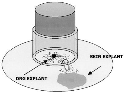
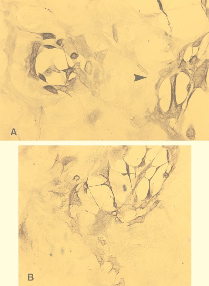
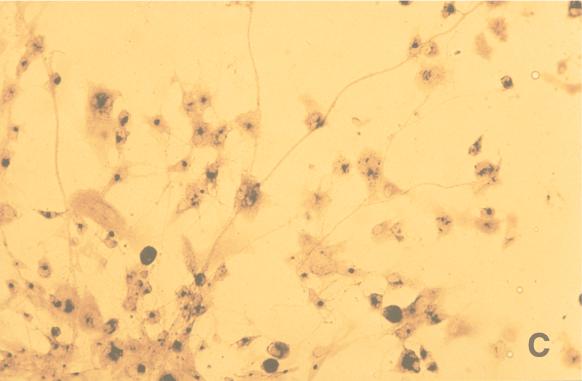
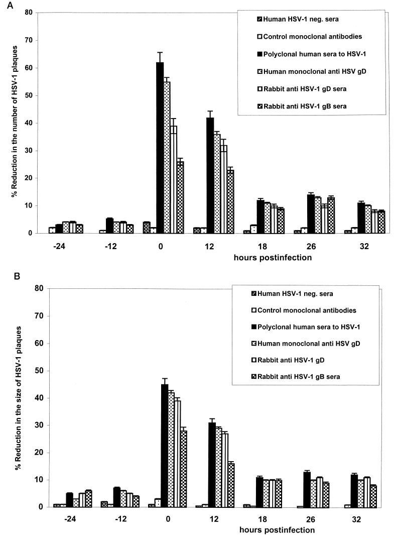
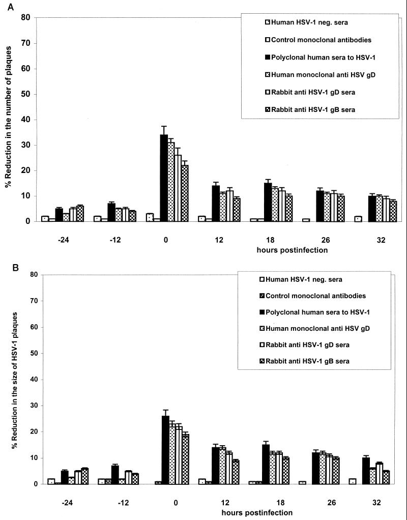
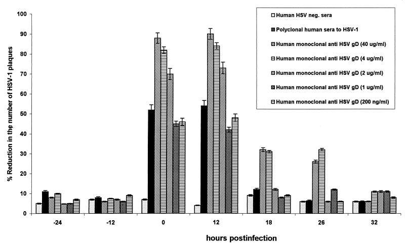
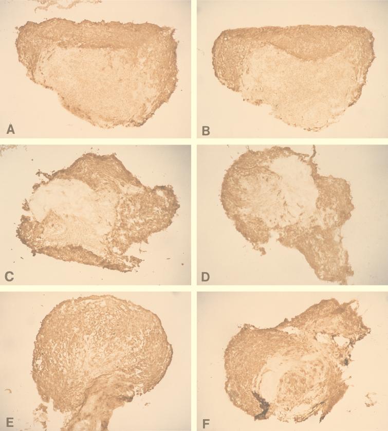
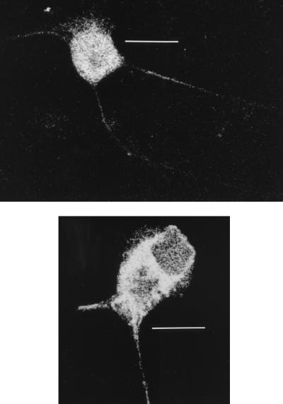
The ability of antibodies to interfere with anterograde transmission of herpes simplex virus (HSV) from neuronal axons to the epidermis was investigated in an in vitro model consisting of human fetal dorsal root ganglia innervating autologous skin explants in a dual-chamber tissue culture system. The number and size of viral cytopathic plaques in epidermal cells after axonal transmission from HSV type 1 (HSV-1)-infected dorsal root ganglionic neurons were significantly reduced by addition to the outer chamber of neutralizing polyclonal human sera to HSV-1, of a human recombinant monoclonal group Ib antibody to glycoprotein D (gD), and of rabbit sera to HSV-1 gB and gD but not by rabbit anti-gE or anti-gG. A similar pattern of inhibition of direct infection of epidermal cells by these antibodies was observed. High concentrations of the monoclonal anti-gD reduced transmission by 90%. Rabbit anti-gB was not taken up into neurons, and human anti-gD did not influence spread of HSV in the dorsal root ganglia or axonal transport of HSV antigens when applied to individual dissociated neurons. These results suggest that anti-gD and -gB antibodies interfere with axonal spread of HSV-1, possibly by neutralizing HSV during transmission across an intercellular gap between axonal termini and epidermal cells, and thus contribute to control of HSV spread and shedding. Therefore, selected human monoclonal antibodies to protective epitopes might even be effective in preventing epidermis-to-neuron transmission during primary HSV infection, especially neonatal infection.


Figures

FIG. 1
FIG. 1
Diagram of the fetal human DRG-EC model.
FIG. 2
FIG. 2
(A and B) Small and large (arrowhead) (A) and medium (B) cytopathic plaques produced by HSV-1 infection in ECs after axonal transmission in the DRG-EC model. Magnification ×320. (C) HSV-1-infected ECs in the outer chamber. Magnification, ×100. Staining was for HSV-1 gC antigen by the immunoperoxidase technique.
FIG. 2
FIG. 2
(A and B) Small and large (arrowhead) (A) and medium (B) cytopathic plaques produced by HSV-1 infection in ECs after axonal transmission in the DRG-EC model. Magnification ×320. (C) HSV-1-infected ECs in the outer chamber. Magnification, ×100. Staining was for HSV-1 gC antigen by the immunoperoxidase technique.
FIG. 3
FIG. 3
Effect of neutralizing and control antibodies on the number (A) and size (B) of cytopathic plaques induced by HSV-1 in ECs after axonal transmission in the DRG-EC model. The HSV inoculum was aspirated after 1 h of incubation, and the DRG in the inner chamber of the model were carefully washed once with HBSS. The antibodies (or growth medium) were incubated for 2 h with ECs in the outer chamber of the model at 24 and 12 h before and 0, 12, 18, 26, and 32 h after infection of the DRG neurons in the inner chamber at the optimal neutralizing dilution. MOI for HSV inoculum, 0.005 TCID50/neuron. neg., negative. Error bars show SEs.
FIG. 4
FIG. 4
Effect of neutralizing and control antibodies on the number (A) and size (B) of cytopathic plaques induced by HSV-1 in ECs after direct infection of the EC monolayers. Monolayers of autologous ECs were obtained by treating the epidermal explants grown to 90% confluence with 0.25% trypsin-EDTA solution (CSL) in HBSS for 2 min at 37°C, washed by centrifugation (800 × g for 7 min), resuspended in growth medium containing 9% FCS, and seeded as a single cells in 12-well plates (Nunc). Twenty-four hours later the autologous ECs were infected at 0.001 and 0.005 TCID50/EC (to approximate the low MOI for EC infection in the DRG-EC model) and treated with antibodies at the same time points as in the DRG-EC model. The cells were washed twice with HBSS after incubation with antibody or HSV-1 infection, fixed with EM-grade methanol, and then stained by the immunoperoxidase method. MOI for HSV inoculum, 0.005 TCID50/EC. neg., negative. Error bars show SEs.
FIG. 5
FIG. 5
Effect of high concentrations of neutralizing human anti-gD monoclonal antibody on the number of cytopathic plaques induced by HSV-1 in ECs in the DRG-EC model. One, 2, 4, or 40 μg of antibody per ml was added to the terminal axons and ECs in the outer chamber at 24 and 12 h before and 0, 12, 24, and 36 h after infection, and the antibody was maintained at this concentration for up to 48 h after infection. After fixation with EM-grade methanol for 10 min, the cultures were stained for gC antigen by immunoperoxidase staining as described in Materials and Methods. MOI for HSV inoculum 0.005 TCID50/neuron. neg., negative. Error bars show SEs.
FIG. 6
FIG. 6
Sections through DRG (isolated from the thoracic region) from HSV-infected DRG-EC cultures at 36 (A and B), 48 (C and D), and 72 (E and F) hpi stained for HSV gC antigen. Human anti-gD (A, C, and E) or control medium (B, D, and F) was added to the outer chamber immediately after infection and left for 12 h. At 36, 48, and 72 hpi, snap-frozen (for 30 s in liquid nitrogen) HSV-infected and mock-infected DRG were mounted on a freezing cryotome (Shandon E-600) at −20°C and sectioned (perpendicularly to the coverslip) into 5-μm-thick sections. The sections were air dried on glass slides, stained with murine anti-gC1 antibody (1:100) (Goodwin Institute for Cancer Research) for 45 min, washed with HBSS and double-distilled water, and stained with biotinylated sheep anti-mouse antibody (Biosource International) (dilution, 1:200) for 45 min at RT. After two washes, sections were treated with streptavidin-horseradish peroxidase conjugate (Biosource International) at a 1:4,000 dilution. The proportion of all DRG neurons which were gC antigen positive was quantified in frozen sections of the whole mounted DRG (as described in Materials and Methods). Actual size of frozen DRG, 1.2 to 2.2 mm.
FIG. 7
FIG. 7
Confocal micrographs of HSV-infected neurons stained for gC antigen at 15 hpi and after addition of human anti-gD monoclonal antibody (top) or control medium (bottom). The HSV inoculum (5 TCID50/cell) was aspirated after 1 h of incubation, and the cells were carefully washed once with HBSS. The HSV-infected or mock-infected dissociated neuronal cultures, incubated with a 1:2,500 dilution (400 ng/ml) of human anti-gD antibody, were fixed in 2.5% formaldehyde (ProSci Tech) in Sorensons buffer (pH 7.4) for 30 min and permeabilized with 0.1% Triton X-100 (Sigma) in PBS for 20 min. Nonspecific staining was blocked by incubation with 5% mouse serum in HBSS for 15 min. The cells on coverslips were then incubated with fluorescein isothiocyanate-conjugated anti-gC1 antibody (Syva Microtrak) (1:100 dilution), rinsed three times with HBSS, and mounted in mounting fluid (Syva Microtrak). Stained neurons were examined with a Bio-Rad MRC 600 confocal microscope. Note the similar distributions of gC antigen in the axon and cytoplasm in both micrographs. Bars, 40 μm (top) and 20 μm (bottom).

Similar articles

Cited by

References

    1. Biron C A, Byron K S, Sullivan J L. Severe herpesvirus infections in an adolescent without natural killer cells. N Engl J Med. 1989;320:1731–1735. - PubMed
    1. Brown Z A, Benedetti J, Ashley R, Burchett S, Selke S, Berry S, Vontver L A, Corey L. Neonatal herpes simplex virus infection in relation to asymptomatic maternal infection at the time of labor. N Engl J Med. 1991;324:1247–1252. - PubMed
    1. Burioni R, Williamson R A, Sanna P P, Bloom F E, Burton D R. Recombinant human Fab to glycoprotein D neutralizes infectivity and prevents cell-to-cell transmission of herpes simplex viruses 1 and 2 in vitro. Proc Natl Acad Sci USA. 1994;91:355–359. - PMC - PubMed
    1. Burlet A J, Menzaghi F, Tilders F J, Oers J W, Nicholas J P, Burlet C R. Uptake of monoclonal antibody to corticotropin-releasing factor (CRF) into rat hypothalamic neurons. Brain Res. 1990;28:283–293. - PubMed
    1. Corey L, Adams H G, Brown Z A, Holmes K K. Genital herpes simplex virus infection: clinical manifestations, course and complications. Ann Intern Med. 1983;98:958–972. - PubMed

Publication types

MeSH terms

Substances

LinkOut - more resources