. 1999 Jul;73(7):5994-6005.
doi: 10.1128/JVI.73.7.5994-6005.1999.

Receptor-mediated Moloney murine leukemia virus entry can occur independently of the clathrin-coated-pit-mediated endocytic pathway

Affiliations

Receptor-mediated Moloney murine leukemia virus entry can occur independently of the clathrin-coated-pit-mediated endocytic pathway

S Lee et al. J Virol. 1999 Jul.

Abstract

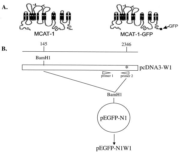
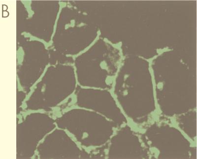
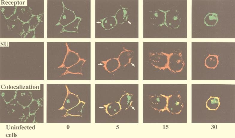
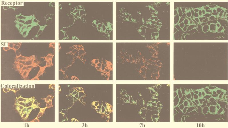
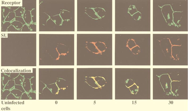
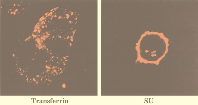
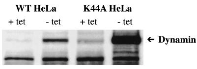
To investigate receptor-mediated Moloney murine leukemia virus (MoMuLV) entry, the green fluorescent protein (GFP)-tagged ecotropic receptor designated murine cationic amino acid transporter (MCAT-1) (MCAT-1-GFP) was constructed and expressed in 293 cells (293/MCAT-1-GFP). 293/MCAT-1-GFP cells displayed green fluorescence primarily at the cell membrane and supported wild-type levels of MoMuLV vector binding and transduction. Using immunofluorescence labeling and confocal microscopy, it was demonstrated that the surface envelope protein (SU) gp70 of MoMuLV virions began to appear inside cells 5 min after virus binding and was colocalized with MCAT-1-GFP. However, clathrin was not colocalized with MCAT-1-GFP, suggesting that MoMuLV entry, mediated by MCAT-1, does not involve clathrin. Double immunofluorescence labeling of SU and clathrin in 293 cells expressing untagged receptor (293/MCAT-1) gave the same results, i.e., SU and clathrin did not colocalize. In addition, we examined the transduction ability of MoMuLV vector on HeLa cells overexpressing the dominant-negative GTPase mutant of dynamin (K44A). HeLa cells overexpressing mutant dynamin have a severe block in endocytosis by the clathrin-coated-pit pathway. No significant titer difference was observed when MoMuLV vector was tranduced into HeLa cells overexpressing either wild-type or mutant dynamin, while the transduction ability of vesicular stomatitis virus glycoprotein pseudotyped vector into HeLa cells overexpressing mutant dynamin was decreased significantly. Taken together, these data suggest that MoMuLV entry does not occur through the clathrin-coated-pit-mediated endocytic pathway.


Figures

FIG. 1
FIG. 1
(A) Schematic representations of the receptor proteins. MCAT-1, ecotropic murine leukemia virus receptor; MCAT-1-GFP, GFP-tagged ecotropic murine leukemia virus receptor. (B) Schematic diagram of the construction of pEGFP-N1W1 (MCAT-1-GFP). pcDNA3-W1 is an MCAT-1 expression plasmid; pEGFP-N1 is a GFP expression plasmid; primer 1, GGCTTTTTACCGGTAGCCGAG; primer 2, CAACCGCTGTCACCCTGGTGGGTGGCCGTGCACGCGGATCCGCTTTGCACTGGTCCAAGTTGC (underlining indicates the BamHI site). ∗, stop codon for MCAT-1. Numbers along the map indicate amino acid positions on MCAT-1. Selected restriction enzyme sites are indicated.
FIG. 2
FIG. 2
Functional analysis of MCAT-1 and MCAT-1-GFP. (A) 293, 293/MCAT-1, and 293/MCAT-1-GFP cells were incubated with MoMuLV at 4°C for 2 h. Binding ability of MCAT-1 and MCAT-1-GFP with MoMuLV was measured by FACS analysis after virus binding followed by incubation with anti-SU monoclonal antibody (83A25) and then R-PE-conjugated goat anti-rat IgG secondary antibody. Transduction ability was assayed on 293/MCAT-1 and 293/MCAT-1-GFP cells by measuring nuclear β-galactosidase activity after transducing cells with an ecotropic retroviral vector containing a nuclear β-galactosidase gene (Ψ2nβg#9). The titer on 3T3 cells was 4 × 10⁶ CFU/ml. (B) 293/MCAT-1-GFP cells showed green fluorescence primarily at the cell surface under a fluorescence microscope.
FIG. 2
FIG. 2
Functional analysis of MCAT-1 and MCAT-1-GFP. (A) 293, 293/MCAT-1, and 293/MCAT-1-GFP cells were incubated with MoMuLV at 4°C for 2 h. Binding ability of MCAT-1 and MCAT-1-GFP with MoMuLV was measured by FACS analysis after virus binding followed by incubation with anti-SU monoclonal antibody (83A25) and then R-PE-conjugated goat anti-rat IgG secondary antibody. Transduction ability was assayed on 293/MCAT-1 and 293/MCAT-1-GFP cells by measuring nuclear β-galactosidase activity after transducing cells with an ecotropic retroviral vector containing a nuclear β-galactosidase gene (Ψ2nβg#9). The titer on 3T3 cells was 4 × 10⁶ CFU/ml. (B) 293/MCAT-1-GFP cells showed green fluorescence primarily at the cell surface under a fluorescence microscope.
FIG. 3
FIG. 3
Colocalization of SU and MCAT-1-GFP in 293/MCAT-1-GFP cells after MoMuLV binding and following incubation at 37°C for different time periods. 293/MCAT-1-GFP cells were incubated with MoMuLV at 37°C for 0, 5, 15, or 30 min. Then cells were fixed, permeabilized, and stained with anti-SU (83A25), biotinylated goat anti-rat IgG secondary antibody, and Cy3-conjugated streptavidin. Color photomicrographs were produced with a Sony printer connected to the video output of the Zeiss confocal microscope. Arrows indicate significant membrane disturbance.
FIG. 4
FIG. 4
Colocalization of SU and MCAT-1-GFP in 293/MCAT-1-GFP cells after MoMuLV binding and following incubation at 37°C for different time periods. 293/MCAT-1-GFP cells were incubated with MoMuLV at 37°C for 1, 3, 7, or 10 h. Cells were then fixed, permeabilized, and stained with anti-SU (83A25), biotinylated goat anti-rat IgG secondary antibody, and Cy3-conjugated streptavidin. Color photomicrographs were produced with a Sony printer connected to the video output of the Zeiss confocal microscope.
FIG. 5
FIG. 5
Colocalization of SU and MCAT-1-GFP in 293/MCAT-1-GFP cells using purified viral particles after binding and following incubation at 37°C for different time periods. 293/MCAT-1-GFP cells were incubated with purified viral particles at 37°C for 0, 5, 15, or 30 min. Then cells were fixed, permeabilized, and stained with anti-SU (83A25), biotinylated goat anti-rat IgG secondary antibody, and Cy3-conjugated streptavidin. Color photomicrographs were produced with a Sony printer connected to the video output of the Zeiss confocal microscope.
FIG. 6
FIG. 6
Indirect immunofluorescence labeling of clathrin and confocal analysis of 293/MCAT-1-GFP cells after MoMuLV binding and incubation at 37°C for different time periods. 293/MCAT-1-GFP cells were bound with MoMuLV vector and incubated at 37°C for 0, 5, 15, or 30 min. Then cells were fixed, permeabilized, and stained with anti-clathrin monoclonal antibody (CHC 5.9), biotinylated goat anti-mouse IgM (μ chain specific) and Cy3-conjugated streptavidin. Color photomicrographs were produced with a Sony printer connected to the video output of the Zeiss confocal microscope. Arrows indicate significant membrane disturbance.
FIG. 7
FIG. 7
Double immunofluorescence labeling of clathrin and SU in 293/MCAT-1 cells after MoMuLV vector binding and incubation at 37°C for different time periods. 293/MCAT-1 cells were bound with MoMuLV and incubated at 37°C for 0, 5, 15, or 30 min. Cells were fixed, permeabilized, and stained with anti-SU (83A25), biotinylated goat anti-rat IgG, Cy3-conjugated streptavidin for SU stain, and then with anti-clathrin monoclonal antibody (CHC 5.9), followed by goat FITC-mouse IgM (μ chain specific) for clathrin staining. Color photomicrographs were produced with a Sony printer connected to the video output of the Zeiss confocal microscope.
FIG. 8
FIG. 8
Transferrin internalization in 293/MCAT-1-GFP cells. 293/MCAT-1-GFP cells were bound with tetramethylrhodamine-conjugated transferrin and incubated at 37°C for 30 min. Cells were fixed, and color photomicrographs were produced with a Sony printer connected to the video output of the Zeiss confocal microscope. (Left panel) Transferrin internalization at 37°C for 30 min. (Right panel) SU staining after virus incubation with 293/MCAT-1-GFP cells at 37°C for 30 min.
FIG. 9
FIG. 9
Western analysis of the wild-type and the dominant-negative GTPase mutant of dynamin (K44A) by inducible expression in stably transformed HeLa cells. Wild-type dynamin-overexpressing HeLa cells (WT HeLa) and dominant-negative GTPase mutant of dynamin-overexpressing HeLa cells (K44A HeLa) were cultured in the presence (+ tet) or absence (− tet) of tetracycline for 50 h to suppress or induce dynamin expression, respectively. Cell lysates were separated on SDS–11 to 14% polyacrylamide gel under reducing conditions. After transfer to Immobilon-P, the blot was probed with anti-HA-peroxidase.

References

    1. Albritton L M, Tseng L, Scadden D, Cunningham J M. A putative murine ecotropic retrovirus receptor gene encodes a multiple membrane-spanning protein and confers susceptibility to virus infection. Cell. 1989;57:659–666. - PubMed
    1. Andersen K B, Nexø B A. Entry of murine retrovirus into mouse fibroblasts. Virology. 1983;125:85–98. - PubMed
    1. Battini J L, Heard J M, Danos O. Receptor choice determinants in the envelope glycoproteins of amphotropic, xenotropic, and polytropic murine leukemia viruses. J Virol. 1992;66:1468–1475. - PMC - PubMed
    1. Berkowitz R D, Goff S P. Point mutations in Moloney murine leukemia virus envelope protein: effects on infectivity, virion association, and superinfection resistance. Virology. 1993;196:748–757. - PubMed
    1. Bruder G, Wiedenmann B. Identification of a distinct 9S form of soluble clathrin in cultured cells and tissues. Exp Cell Res. 1986;164:449–462. - PubMed

Publication types

MeSH terms

LinkOut - more resources