. 1999 Jul;73(7):6015-23.
doi: 10.1128/JVI.73.7.6015-6023.1999.

Hepatitis A virus capsid protein VP1 has a heterogeneous C terminus

Affiliations

Hepatitis A virus capsid protein VP1 has a heterogeneous C terminus

J Graff et al. J Virol. 1999 Jul.

Abstract

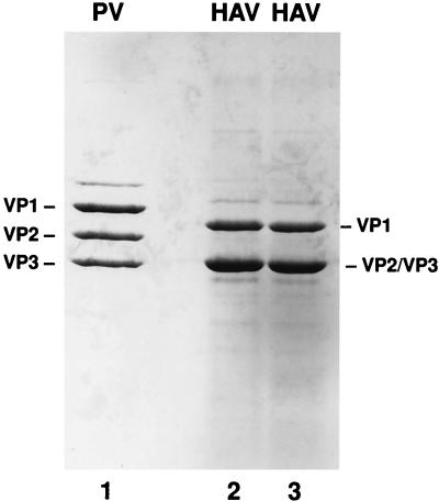
Hepatitis A virus (HAV) encodes a single polyprotein which is posttranslationally processed into the functional structural and nonstructural proteins. Only one protease, viral protease 3C, has been implicated in the nine protein scissions. Processing of the capsid protein precursor region generates a unique intermediate, PX (VP1-2A), which accumulates in infected cells and is assumed to serve as precursor to VP1 found in virions, although the details of this reaction have not been determined. Coexpression in transfected cells of a variety of P1 precursor proteins with viral protease 3C demonstrated efficient production of PX, as well as VP0 and VP3; however, no mature VP1 protein was detected. To identify the C-terminal amino acid residue of HAV VP1, we performed peptide sequence analysis by protease-catalyzed [18O]H2O incorporation followed by liquid chromatography ion-trap microspray tandem mass spectrometry of HAV VP1 isolated from purified virions. Two different cell culture-adapted isolates of HAV, strains HM175pE and HM175p35, were used for these analyses. VP1 preparations from both virus isolates contained heterogeneous C termini. The predominant C-terminal amino acid in both virus preparations was VP1-Ser274, which is located N terminal to a methionine residue in VP1-2A. In addition, the analysis of HM175pE recovered smaller amounts of amino acids VP1-Glu273 and VP1-Thr272. In the case of HM175p35, which contains valine at amino acid position VP1-273, VP1-Thr272 was found in addition to VP1-Ser274. The data suggest that HAV 3C is not the protease responsible for generation of the VP1 C terminus. We propose the involvement of host cell protease(s) in the production of HAV VP1.


Figures

FIG. 1
FIG. 1
Schematic representation of subgenomic HAV constructs used to examine the capsid protein cleavage mediated by HAV 3C. The constructs encode various lengths of HAV protein sequences up to the nucleotide position indicated on the right of each HAV diagram, downstream of the EMCV IRES. All plasmids contain a T7 RNA polymerase promoter upstream of the EMCV sequence. The predicted molecular mass (MW) of each uncleaved protein is indicated to the right. A diagram of the HAV polyprotein is shown on top for comparison.
FIG. 2
FIG. 2
Immunoblot analysis of HAV polyproteins expressed from cDNA in the presence of HAV 3C in BS-C-1 cells. Transient transfection was performed in the presence of recombinant vaccinia virus vTF7-3, which carries the gene for T7 RNA polymerase. Proteins P1-2380, P1-2993, and P1P2 were expressed from plasmids pEHP1-2380, pEHP1-2993, and pEHP1P2, respectively, in the absence (A and C, lanes 3, 5, and 7) or presence (A and C, lanes 4, 6, and 8) of HAV 3C expressed from pE5H-P3. VP1-specific products were detected with HAV VP1 antiserum (A), and VP4-specific products were detected with HAV VP4 antiserum (C). (B) VP1-immunoreactive proteins P1-3107 and P1-3216 expressed from plasmids pEHP1-3107 and pEHP1-3216, respectively, in the absence (lanes 3 and 5) or presence (lanes 4 and 6) of HAV 3C expressed from pE5H-P3. Extract from mock-transfected BS-C-1 cells as a negative control (NC) and extract from HAV-infected cells as a positive control (HAV) were analyzed on the same gel (all panels). Immunoreactive proteins VP1 (A and B), PX (A), and VP0VP3 and VP0 (C) are indicated to the right; molecular masses of marker proteins are indicated in kilodaltons to the left (all panels).
FIG. 3
FIG. 3
Polyacrylamide-SDS gel analysis of HAV capsid proteins. HM175p35 was propagated in 11-1 cells and purified by CsCl isopycnic gradient centrifugation. Capsid proteins were separated by SDS-PAGE (lanes 2 and 3) in comparison with PV (lane 1). The gel was stained with Coomassie blue for excision of capsid protein VP1. PV capsid proteins are indicated to the left, and HAV capsid proteins are indicated to the right.
FIG. 4
FIG. 4
Principle of 18O incorporation into the internal α-carboxyl group upon proteolytic cleavage of the polypeptide chain. In the presence of 50% 18O-enriched buffer, 18O incorporation is excluded from the C-terminal peptide.
FIG. 5
FIG. 5
LC-MS/MS analysis of capsid protein VP1. A reverse-phase chromatogram of the resulting peptides derived from the tryptic digests of PV VP1 (A) and HAV VP1 (B) is shown. The asterisks and bar indicate the regions which identified the C-terminal peptides in further analyses.
FIG. 6
FIG. 6
Mass spectral analysis of individual peptide peaks from the reverse-phase chromatogram of PV VP1 (Fig. 5A). The ion signals of two representative peptides for which 50% of the molecules have incorporated 18O are shown ([M−H]+, 456.2 [A] and 1,032.5 [B]). The isotope distribution in panels A and B indicates the mixture of two peptide species, one containing 16O (peaks of 456.2 and 1,032.5); the other species has exchanged one 16O for one 18O resulting in a net mass increase by two mass units for 50% of the molecules (peaks of 458.2 and 1,034.5). In contrast, the isotope distribution of the [M−H]+ ion at m/z 612.1 reveals no 18O incorporation (C) and therefore is the carboxy-terminal peptide.
FIG. 7
FIG. 7
MS/MS analysis of the putative C-terminal peptide of PV VP1. The MS/MS spectrum of the [M−H]+ ion at m/z 612.1 was generated during the LC-MS/MS analysis of the PV VP1 tryptic digestion mixture. The fragmentation pattern reveals the C-terminal peptide expected sequence. The b-ions are NH2-terminal fragments derived from successive removal of amino acids from the COOH terminus, and the y-ions are COOH-terminal fragments derived from successive removal of amino acids from the NH2 terminus.
FIG. 8
FIG. 8
MS/MS analysis of the putative C-terminal peptides of HAV VP1 derived from HM175pE. Three ion signals corresponding to C-terminal peptides of HAV VP1 were identified. The isotopic distribution of the doubly charged ions is shown in the insets of panels A, B, and C. The sequences were derived by MS/MS analysis. The mass difference of 87 amu of the y-ion series generated from the peptide in panel A to the y-ion series generated from the peptide in panel B denotes the lack of a serine residue in the peptide (marked in red). The mass difference of 129 amu similarly observed during the CID analysis of the peptides displayed in panels B and C denotes loss of a glutamate residue in panel C (marked in green).
FIG. 9
FIG. 9
C-terminal sequence of capsid protein VP1 determined by trypsin-catalyzed 18O incorporation followed by LC-MS/MS. An asterisk indicates that oxidation of the methionine residue was detected in all HAV VP1 peptides. The predominant C-terminal amino acid (aa) of HAV VP1 detected in both cases was Ser274.

References

    1. Anderson D A, Ross B C. Morphogenesis of hepatitis A virus: isolation and characterization of subviral particles. J Virol. 1990;64:5284–5289. - PMC - PubMed
    1. Baily J M, Shenoy N R, Ronk M, Shively J E. Automated carboxy-terminal sequence analysis of peptides. Protein Sci. 1992;1:68–80. - PMC - PubMed
    1. Bergmann E M, Mosimann S C, Chernaia M M, Malcolm B A, James M N G. The refined crystal structure of the 3C gene product from hepatitis A virus: specific proteinase activity and RNA recognition. J Virol. 1997;71:2436–2448. - PMC - PubMed
    1. Bishop N E, Anderson D A. Hepatitis A virus subviral particles: purification, accumulation, and relative infectivity of virions, provirions and procapsids. Arch Virol. 1997;142:2147–2160. - PubMed
    1. Boege U, Scraba D G. Mengo virus maturation is accompanied by C-terminal modification of capsid protein VP1. Virology. 1989;168:409–412. - PubMed

Publication types

MeSH terms

LinkOut - more resources