. 1999 Jul;73(7):6104-10.
doi: 10.1128/JVI.73.7.6104-6110.1999.

Dengue virus type 1 nonstructural glycoprotein NS1 is secreted from mammalian cells as a soluble hexamer in a glycosylation-dependent fashion

Affiliations

Dengue virus type 1 nonstructural glycoprotein NS1 is secreted from mammalian cells as a soluble hexamer in a glycosylation-dependent fashion

M Flamand et al. J Virol. 1999 Jul.

Abstract

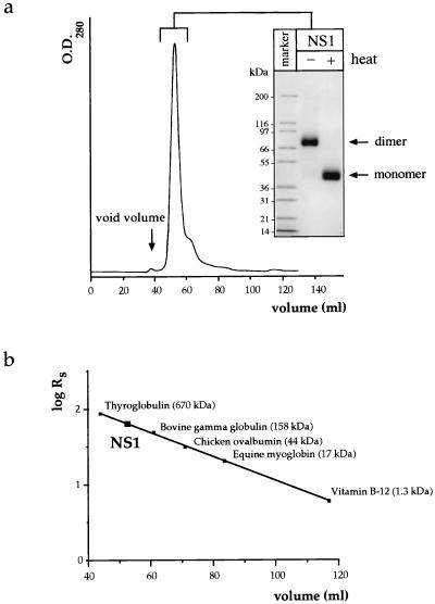
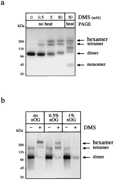
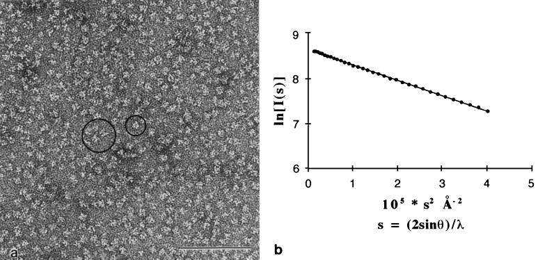
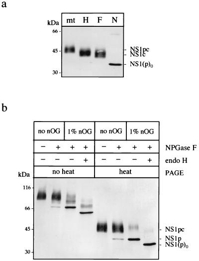
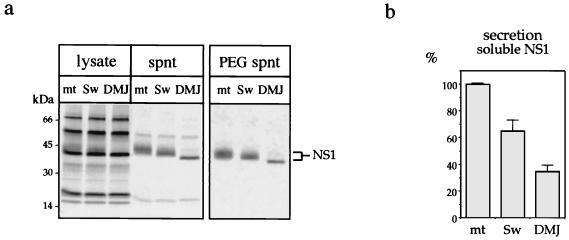
Nonstructural glycoprotein NS1, specified by dengue virus type 1 (Den-1), is secreted from infected green monkey kidney (Vero) cells in a major soluble form characterized by biochemical and biophysical means as a unique hexameric species. This noncovalently bound oligomer is formed by three dimeric subunits and has a molecular mass of 310 kDa and a Stokes radius of 64.4 A. During protein export, one of the two oligosaccharides of NS1 is processed into an endo-beta-N-acetylglucosaminidase F-resistant complex-type sugar while the other remains of the polymannose type, protected in the dimeric subunit from the action of maturation enzymes. Complete processing of the complex-type sugar appears to be required for efficient release of soluble NS1 into the culture fluid of infected cells, as suggested by the repressive effects of the N-glycan processing inhibitors swainsonine and deoxymannojyrimicin. These results, together with observations related to the absence of secretion of NS1 from Den-infected insect cells, suggest that maturation and secretion of hexameric NS1 depend on the glycosylation status of the host cell.


Figures

FIG. 1
FIG. 1
Analysis of immunoaffinity-purified soluble extracellular NS1 by SEC. (a) Immunoaffinity-purified NS1 was submitted to SEC on an S300 gel filtration column, and the single peak was concentrated by ultrafiltration and analyzed on a 4 to 20% gradient SDS-polyacrylamide gel stained by Coomassie blue. The protein was either unheated or heated to 95°C for 3 min prior to electrophoresis. (b) By comparison with protein standards used to calibrate the SEC column, the NS1 protein exhibited a Stokes radius (RS) of 64.4 Å, corresponding to an apparent molecular mass of 300 kDa.
FIG. 2
FIG. 2
Purified extracellular NS1 is a hexamer that can be converted to its dimeric subunit in the presence of the nonionic detergent nOG, as demonstrated by chemical cross-linking. (a) Subsequent to SEC, the protein was concentrated to 0.5 mg/ml by ultrafiltration and treated with final concentrations of 0, 0.5, 5, and 50 mM DMS. The resulting products were placed in nonreducing Laemmli sample buffer, separated on a 4 to 20% gradient acrylamide gel, and stained with Coomassie blue. One sample, treated with 50 mM DMS, was treated for 3 min at 95°C prior to electrophoresis to dissociate noncovalently linked oligomers. (b) Purified NS1 was treated overnight at 37°C with 0, 0.5, or 1% nOG and submitted or not submitted to 25 mM DMS for 1 h. Proteins were separated without heat denaturation on a 4 to 20% gradient acrylamide gel and detected by immunoblotting with MAb 3D1.4.
FIG. 3
FIG. 3
Hexameric NS1 appears as a unique species by electron microscopy and SAXS analysis. (a) Negatively stained sample of purified NS1 at 0.2 mg/ml observed by electron microscopy at 80 kV. The image shows a field containing NS1 particles in random orientations. Dimensions are quite homogeneous, with a diameter of roughly 11 nm. Occasionally, particles lying with a twofold (large circle) or a threefold (small circle) axis of symmetry perpendicular to the plane of the figure are seen. Bar, 0.1 μm. (b) Guinier plot of purified NS1 at a protein concentration of 6.2 mg/ml. The radius of gyration (see text) calculated from the slope, Rg, is 5 ± 0.1 nm. Note that the curve is linear from very small angles (lower s values), showing that there is a single form of NS1 protein in solution. The molecular mass estimated from extrapolation to s = 0 is 300 ± 50 kDa.
FIG. 4
FIG. 4
Analysis of the nature and accessibility of the two oligosaccharides of extracellular NS1. (a) Extracellular NS1 was mock treated (mt) or treated for 1 h at 37°C with endo H (H), endo F (F), or NPGase F (N) in its monomeric form. Samples were placed in nonreducing Laemmli sample buffer and separated by SDS–12% PAGE, and the resulting products were detected by immunoblotting after transfer to nitrocellulose. “p” indicates the presence of a polymannose-type oligosaccharide; “c” indicates a complex-type oligosaccharide on the protein. NS1(p)0 is the deglycosylated form of the protein. (b) Purified NS1 was either mock treated or treated with NPGase F for 1 h at 37°C in 10 mM sodium phosphate buffer, pH 7.5. One sample was further treated by endo H after the pH was readjusted to 5.5 with 50 mM citrate buffer. All samples were subjected to SDS–12% PAGE with or without preliminary heat denaturation in nonreducing Laemmli sample buffer.
FIG. 5
FIG. 5
Altered secretion of NS1 from infected cells treated with N-glycan processing inhibitors. (a) Cells were infected with Den virus at an m.o.i. of 3. At 7 h postinfection, swainsonine (Sw) and DMJ were added for 20 h at concentrations of 5 μM and 1 mM, respectively. Proteins were then radiolabeled for 3.5 h; immunoprecipitated with Den virus-specific PAb from cell lysates, total supernatants (spnt), or PEG-treated supernatants; separated by SDS-PAGE; and analyzed by autoradiography. (b) NS1-related radioactive signals from PEG-treated supernatants immunoprecipitated with the PAb were quantified directly from the gels with a PhophorImager. Average values of at least three independent experiments and corresponding standard deviations are reported on the graph. mt, mock treatment.

References

    1. Bergh M, Cepko C, Wolf D, Robbins P. Expression of the Saccharomyces cerevisiae glycoprotein invertase in mouse fibroblasts: glycosylation, secretion, and enzymatic activity. Proc Natl Acad Sci USA. 1987;84:3570–3574. - PMC - PubMed
    1. Boulin C, Kempf R, Koch M H J, Mclaughlin S M. Data appraisal, evaluation and display for synchrotron radiation experiments: hardware and software. Nucl Instrum Methods. 1986;A249:399–407.
    1. Chambers T J, Hahn C S, Galler R, Rice C M. Flavivirus genome organization, expression, and replication. Annu Rev Microbiol. 1990;44:649–688. - PubMed
    1. Crooks A J, Lee J M, Dowsett A B, Stephenson J R. Purification and analysis of infectious virions and native non-structural antigens from cells infected with tick-borne encephalitis virus. J Chromatogr. 1990;502:59–68. - PubMed
    1. Crooks A J, Lee J M, Easterbrook L M, Timofeev A V, Stephenson J R. The NS1 protein of tick-borne encephalitis virus forms multimeric species upon secretion from the host cell. J Gen Virol. 1994;75:3453–3460. - PubMed

Publication types

Substances

LinkOut - more resources