

doi: 10.1128/JVI.73.7.6182-6187.1999.
The subgenus-specific C-terminal region of protein IX is located on the surface of the adenovirus capsid
Affiliations
- PMID: 10364380
- PMCID: PMC112689
- DOI: 10.1128/JVI.73.7.6182-6187.1999
Item in Clipboard
The subgenus-specific C-terminal region of protein IX is located on the surface of the adenovirus capsid
J Virol.
1999 Jul.
Abstract
We have investigated the antigenicity of the C- and N-terminal halves of pIX of human adenovirus types 2 and 3 (Ad2 and Ad3) as well as their orientations in virions. We found that only the C-terminal halves of Ad2 pIX and Ad3 pIX reacted in a subgenus-specific manner by enzyme-linked immunosorbent assay and immunoblot analysis. Based on immunoelectron microscopy experiments, pIX in viral capsids appears to be positioned such that the C-terminal part of pIX constitutes the surface domain whereas the N terminus of the protein makes up the internal domain in icosahedral Ad capsids.
Figures
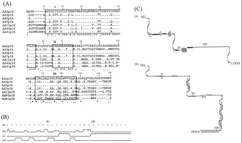
(A) Multiple amino acid sequence alignment of pIX from seven human Ad serotypes. Residue numbering includes the gaps (−) in the alignment. The asterisks and dots denote identical and conserved residues, respectively. Structural similarity is indicated by boxes (labelled I, II, and III). (B) Schematic diagram of the secondary structure derived from predictions made by the Chou and Fasman method (10) shows residue numbering according to the sequences of Ad2 (a), turns (b), alpha-helical regions (c), and β regions (d). (C) Schematic diagrams of the potential antigenic sites of Ad2pIX (a) and Ad3pIX (b) (23). The sites are represented by octagons.
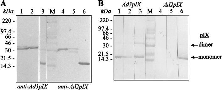
Reactivities of antisera to recombinant full-length pIX and partially deleted pIX demonstrated by immunoblotting. (A) Analysis of reactivities of recombinant full-length pIX and C- and N-terminal halves of pIX with full-length pIX-specific polyclonal rabbit sera. Proteins Ad3pIXC, Ad3pIXN, and Ad3 pIX were electrotransferred to nitrocellulose after denaturing sodium dodecyl sulfate-polyacrylamide gel electrophoresis and incubated with anti-Ad3 pIX as a primary antibody (lanes 1, 2 and 3); Ad2pIXC, Ad2pIXN, and Ad2 pIX were incubated with anti-Ad2 pIX (lanes 4, 5, and 6). Sera were diluted at 1:500. Horseradish peroxidase-conjugated secondary antibody was used with 3,3′-5,5′-tetramethylbenzidine (Boehringer, Mannheim, Germany) as a substrate. (B) Analysis of full-length recombinant Ad3 pIX (lanes 1 to 3) and Ad2 pIX (lanes 4 to 6). The antibodies used were as follows: lane 1, anti-Ad3pIXC; lane 2, anti-Ad3pIXN; lane 3, anti-Ad3 pIX; lane 4, anti-Ad2pIXC; lane 5, anti-Ad2pIXN; lane 6, anti-Ad2 pIX. Lane M, molecular weight markers. The positions of markers are given on the left.
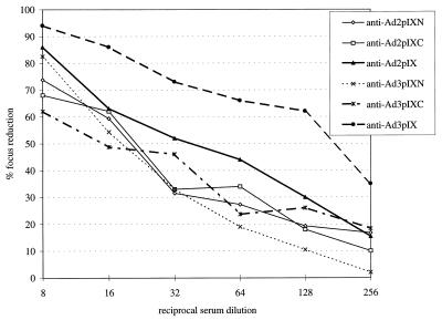
Neutralization of the homologous Ad by polyclonal antisera directed to the full-length Ad2 and Ad3 pIX and to the terminal halves of each pIX. The result for each focus reduction assay is the average of three independent determinations of duplicate samples. Standard deviations were within the range from 5 to 20%.
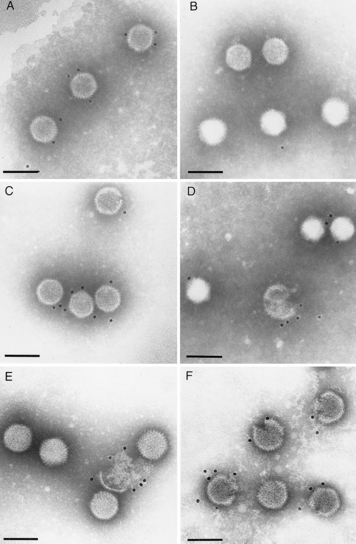
Localization of pIX on capsid surfaces of Ad particles by immunogold electron microscopy performed by using anti-Ad3 pIX serum (A and F), anti-Ad3pIXN serum (B), anti-Ad3pIXC serum (C), and anti-Ad2 pIX serum (D and E). In all panels bars indicate 100 nm.
References
-
- Akalu A. Antigenic characterization and posttranslational modification analysis of the protein pIX of human adenovirus serotypes 2 and 3. Thesis. Greifswald, Germany: University of Greifswald; 1997.
-
- Akalu A, Seidel W, Liebermann H, Bauer U, Döhner L. Rapid identification of subgenera of human adenovirus by serological and PCR assays. J Virol Methods. 1998;71:187–196. - PubMed
-
- Aleström P, Akusjärvi G, Perricaudet M, Mathews M B, Klessig D F, Pettersson U. The gene for polypeptide IX of adenovirus type 2 and its unspliced messenger RNA. Cell. 1980;19:671–681. - PubMed
-
- Allard A, Wadell G. The E1B transcription map of the enteric adenovirus type 41. Virology. 1992;188:319–330. - PubMed
-
- Bos J L, Polder L J, Bernards R, Schrier P I, van der Elsen P J, van der Eb A J, van Ormondt H. The 2.2 kb E1b mRNA of human Ad12 and Ad5 codes for two tumor antigens starting at different AUG triplets. Cell. 1981;27:121–131. - PubMed
Publication types
MeSH terms
Substances
LinkOut - more resources
Full Text Sources
Other Literature Sources
