

Upper respiratory tract disease in the gopher tortoise is caused by Mycoplasma agassizii
- PMID: 10364595
- PMCID: PMC85132
- DOI: 10.1128/JCM.37.7.2262-2269.1999
Upper respiratory tract disease in the gopher tortoise is caused by Mycoplasma agassizii
Abstract
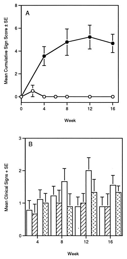
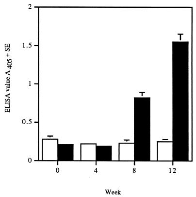
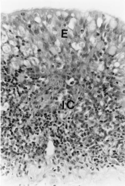
Upper respiratory tract disease (URTD) has been observed in a number of tortoise species, including the desert tortoise (Gopherus agassizii) and the gopher tortoise (Gopherus polyphemus). Clinical signs of URTD in gopher tortoises are similar to those in desert tortoises and include serous, mucoid, or purulent discharge from the nares, excessive tearing to purulent ocular discharge, conjunctivitis, and edema of the eyelids and ocular glands. The objectives of the present study were to determine if Mycoplasma agassizii was an etiologic agent of URTD in the gopher tortoise and to determine the clinical course of the experimental infection in a dose-response infection study. Tortoises were inoculated intranasally with 0.5 ml (0.25 ml/nostril) of either sterile SP4 broth (control group; n = 10) or 10(8) color-changing units (CCU) (total dose) of M. agassizii 723 (experimental infection group; n = 9). M. agassizii caused clinical signs compatible with those observed in tortoises with natural infections. Clinical signs of URTD were evident in seven of nine experimentally infected tortoises by 4 weeks postinfection (p.i.) and in eight of nine experimentally infected tortoises by 8 weeks p.i. In the dose-response experiments, tortoises were inoculated intranasally with a low (10(1) CCU; n = 6), medium (10(3) CCU; n = 6), or high (10(5) CCU; n = 5) dose of M. agassizii 723 or with sterile SP4 broth (n = 10). At all time points p.i. in both experiments, M. agassizii could be isolated from the nares of at least 50% of the tortoises. All of the experimentally infected tortoises seroconverted, and levels of antibody were statistically higher in infected animals than in control animals for all time points of >4 weeks p.i. (P < 0.0001). Control tortoises in both experiments did not show clinical signs, did not seroconvert, and did not have detectable M. agassizii by either culture or PCR at any point in the study. Histological lesions were compatible with those observed in tortoises with natural infections. The numbers of M. agassizii 723 did not influence the clinical expression of URTD or the antibody response, suggesting that the strain chosen for these studies was highly virulent. On the basis of the results of the transmission studies, we conclude that M. agassizii is an etiologic agent of URTD in the gopher tortoise.
Figures
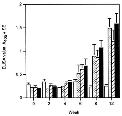
 , low dose; ▨, medium dose; ■, high dose) and
control gopher tortoises (□).
, low dose; ▨, medium dose; ■, high dose) and
control gopher tortoises (□).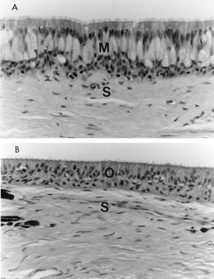
References
-
- Berry K H. Demographic consequences of disease in two desert tortoise populations in California, USA. In: Van Ebbema J, editor. Proceedings: conservation, restoration, and management of tortoises and turtles—an international conference. New York, N.Y: Wildlife Conservation Society Turtle Recovery Program and the New York Turtle and Tortoise Society; 1997. pp. 91–99.
-
- Brown D R, Crenshaw B C, McLaughlin G S, Schumacher I M, McKenna C E, Klein P A, Jacobson E R, Brown M B. Taxonomy of the tortoise mycoplasmas Mycoplasma agassizii and Mycoplasma testudinisby 16S rRNA gene sequence comparisons. Int J Syst Bacteriol. 1995;45:348–350. - PubMed
-
- Brown, D. R., M. B. Brown, and J. G. Tully. Comparison of mycoplasma isolated from alligators and crocodiles, abstr. G-10, p. 281. In Abstracts of the 97th General Meeting of the American Society for Microbiology 1997. American Society for Microbiology, Washington, D.C.
-
- Brown, M. B. Unpublished data.
Publication types
MeSH terms
Substances
LinkOut - more resources
Full Text Sources
