

Case Reports
doi: 10.1128/JCM.37.7.2378-2380.1999.
Chlamydia pneumoniae in a free-ranging giant barred frog (Mixophyes iteratus) from Australia
Affiliations
- PMID: 10364623
- PMCID: PMC85174
- DOI: 10.1128/JCM.37.7.2378-2380.1999
Item in Clipboard
Case Reports
Chlamydia pneumoniae in a free-ranging giant barred frog (Mixophyes iteratus) from Australia
J Clin Microbiol.
1999 Jul.
Abstract
The koala biovar of Chlamydia pneumoniae was identified in lung tissue from a sick, free-ranging giant barred frog (Mixophyes iteratus) by using electron microscopy, C. pneumoniae-specific fluorescent-antibody staining, cell culture, and sequencing of the ompA, ompB and 16S rRNA genes. This is the first report of a chlamydial strain infecting both a homeotherm and a poikilotherm and only the fourth host (in addition to humans, koalas, and horses) to be naturally infected with this species of Chlamydia. The frog had severe, chronic, mononuclear pneumonia and nonregenerative anemia and pancytopenia.
Figures

Histological section of lung tissue from a giant barred frog with severe, chronic, mononuclear pneumonia. The septae, which are normally covered by a thin epithelium, are here markedly thickened by a layer of mononuclear inflammation (arrowheads). H&E stain. Bar, 500 μm.
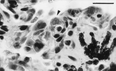
Histological section of lung tissue with an infected mononuclear cell (arrowhead). It is important to note the swollen cytoplasm containing chlamydial organisms, visible as fine stippling. H&E stain. Bar, 20 μm.
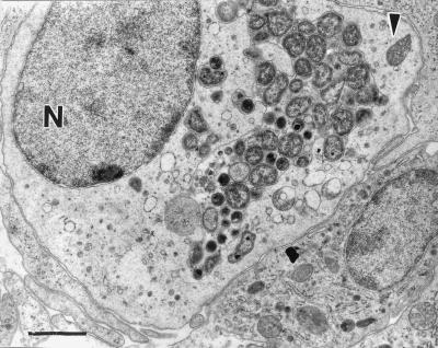
Transmission electron micrograph of infected mononuclear cell with chlamydial particles at various stages present within a membrane-bound cytoplasmic inclusion. N, nucleus. The arrowhead indicates a mitochondrion. Bar, 1,500 nm.
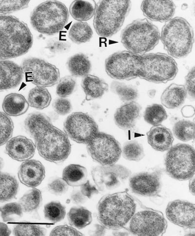
Transmission electron micrograph of chlamydial particles at various stages in lung tissue. It is important to note the large reticulate bodies (R), which undergo binary fission, intermediate bodies (I), and condensed elementary bodies (E). Bar, 700 nm.
References
-
- Cox R, Kuo C-C, Grayston T, Campbell L A. Deoxyribonucleic acid relatedness of Chlamydia sp. strain TWAR to Chlamydia trachomatis and Chlamydia psittaci. Int J Syst Bacteriol. 1988;38:265–268.
-
- Drury R A, Wallington E A. Carleton’s histological technique. Oxford, United Kingdom: Oxford University Press; 1980. p. 264.
-
- Fukushi H, Hirarai K. Proposal of Chlamydia pecorum sp. nov. for Chlamydia strains derived from ruminants. Int J Syst Bacteriol. 1992;42:306–308. - PubMed
Publication types
MeSH terms
Substances
Associated data
- Actions
- Actions
- Actions
LinkOut - more resources
Full Text Sources
Other Literature Sources
Medical
