

Magnetic resonance imaging of the wrist in early rheumatoid arthritis reveals progression of erosions despite clinical improvement
- PMID: 10364913
- PMCID: PMC1752839
- DOI: 10.1136/ard.58.3.156
Magnetic resonance imaging of the wrist in early rheumatoid arthritis reveals progression of erosions despite clinical improvement
Abstract
Objectives: To investigate the progression of joint damage in early rheumatoid arthritis (RA) using magnetic resonance imaging (MRI) of the wrist and determine whether this technique can be used to predict prognosis.
Methods: An inception cohort of 42 early patients has been followed up prospectively for one year. Gadolinium enhanced MRI scans of the dominant wrist were obtained at baseline and one year and scored for synovitis, tendonitis, bone marrow oedema, and erosions. Plain radiographs were performed concurrently and scored for erosions. Patients were assessed clinically for disease activity and HLA-DRB1 genotyping was performed.
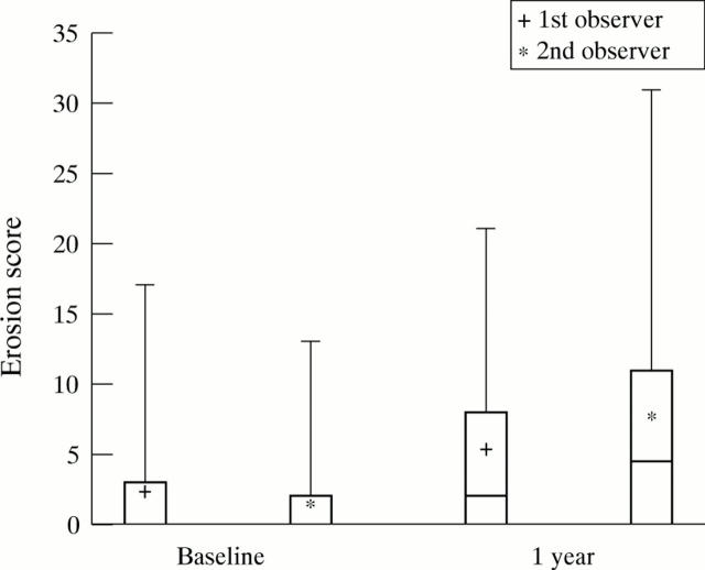
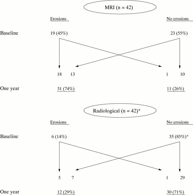
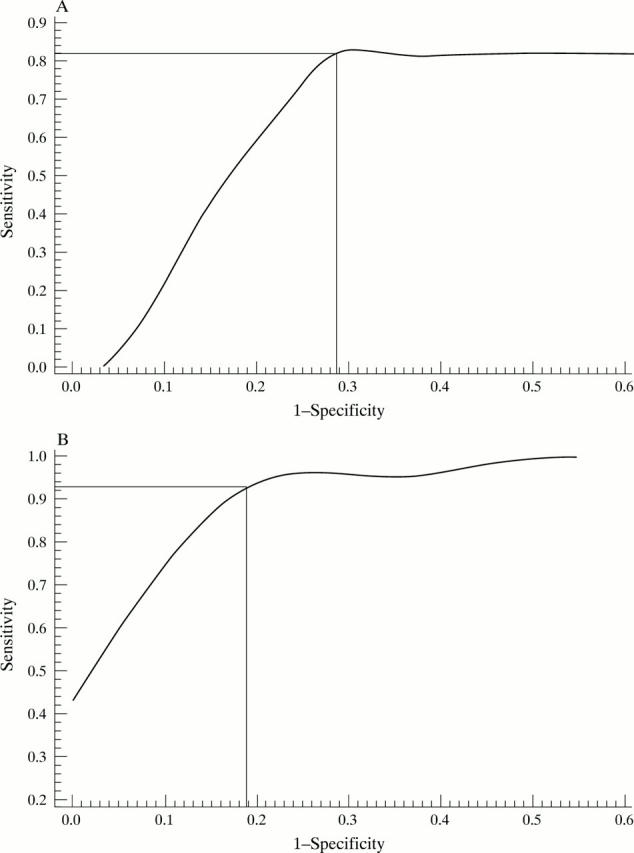
Results: At one year, MRI erosions were found in 74% of patients (31 of 42) compared with 45% at baseline. Twelve patients (28.6%) had radiographic erosions at one year. The total MRI score and MRI erosion score increased significantly from baseline to one year despite falls in clinical measures of inflammation including erythrocyte sedimentation rate (ESR), C reactive protein (CRP), and swollen joint count (p < 0.01 for all). Baseline findings that predicted carpal MRI erosions at one year included a total MRI score of 6 or greater (sensitivity: 93.3%, specificity 81.8%, positive predictive value 93.3%, p = 0.000007), MRI bone oedema (OR = 6.47, p < 0.001), MRI synovitis (OR = 2.14, p = 0.003), and pain score (p = 0.01). Radiological erosions at one year were predicted by a total MRI score at baseline of greater than 13 (OR = 12.4, p = 0.002), the presence of MRI erosions (OR = 11.6, p = 0.005), and the ESR (p = 0.02). If MRI erosions were absent at baseline and the total MRI score was low, radiological erosions were highly unlikely to develop by one year (negative predictive value 0.91 and 0.92 respectively). No association was found between the shared epitope and erosions on MRI (p = 0.4) or radiography (p = 1.0) at one year.
Conclusions: MRI scans of the dominant wrist are useful in predicting MRI and radiological erosions in early RA and may indicate the patients that should be managed aggressively. Discordance has been demonstrated between clinical improvement and progression of MRI erosion scores.
Figures
References
Publication types
MeSH terms
Substances
LinkOut - more resources
Full Text Sources
Medical
Research Materials
Miscellaneous
