. 1999 Jun 15;19(12):4748-54.
doi: 10.1523/JNEUROSCI.19-12-04748.1999.

Characterization of phosphorylation sites on the glutamate receptor 4 subunit of the AMPA receptors

Affiliations

Characterization of phosphorylation sites on the glutamate receptor 4 subunit of the AMPA receptors

A L Carvalho et al. J Neurosci. .

Abstract

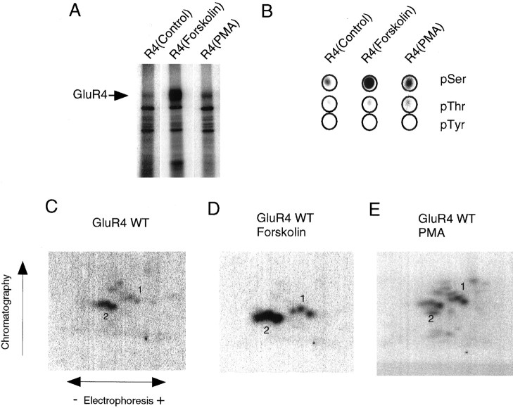
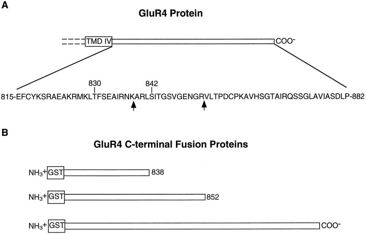
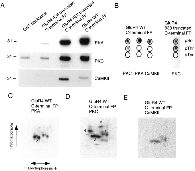
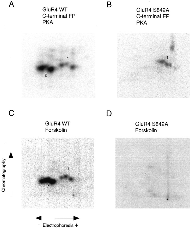
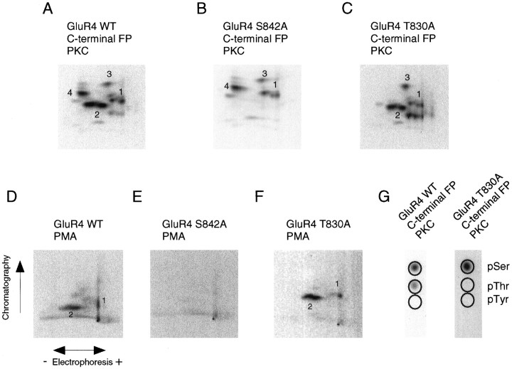
Recent studies have suggested that protein phosphorylation of glutamate receptors may play an important role in synaptic transmission. Specifically, the phosphorylation of AMPA receptors has been implicated in cellular models of synaptic plasticity. The phosphorylation of the glutamate receptor 1 (GluR1) subunit of AMPA receptors by protein kinase A (PKA), protein kinase C (PKC), and Ca2+/calmodulin-dependent protein kinase II (CaMKII) has been characterized extensively. Phosphorylation of this subunit occurs exclusively on the intracellular C-terminal domain. However, the GluR1 subunit C terminus shows low homology to the other AMPA receptor subunits. In this paper we characterized the phosphorylation of AMPA receptor subunit GluR4, using site-specific mutagenesis and biochemical techniques. We found that GluR4 is phosphorylated on serine 842 within the C-terminal domain in vitro and in vivo. Serine 842 is phosphorylated by PKA, PKC, and CaMKII in vitro and is phosphorylated in transfected cells by PKA. Two-dimensional phosphopeptide analysis indicates that serine 842 is the major phosphorylation site on GluR4. In addition, we identified threonine 830 as a potential PKC phosphorylation site. These results suggest that GluR4, which is the most rapidly desensitizing AMPA receptor subunit, may be modulated by phosphorylation.


Figures

Fig. 1.
Fig. 1.
Phosphorylation of GluR4 AMPA receptor subunit transiently expressed in HEK 293T cells. A, GluR4 was immunoprecipitated from labeled cells, resolved by SDS-PAGE, and visualized by autoradiography. B, After digestion with trypsin, GluR4 was hydrolyzed with 6N HCl, and the resulting amino acids were separated by electrophoresis. The circlesindicate the migration of ninhydrin-stained phosphoamino acid standards. GluR4 phosphoamino acids were visualized by autoradiography.C–E, Phosphorylated GluR4 was excised from the gel and digested with trypsin; the resulting phosphopeptides were spotted onto chromatography plates and resolved in two dimensions. HEK 293T cells expressing GluR4 were labeled with 1 mCi/ml [32P] orthophosphate and treated for 15 min with control solution (C), with 10 μm forskolin (D), or with 200 nm PMA (E).
Fig. 2.
Fig. 2.
GluR4 C terminal. A, Schematic diagram of the C-terminal region of GluR4, including the fourth transmembrane domain (TMD IV). Theexpanded region shows the amino acid sequence of the C terminal, and the residues subjected to site-directed mutagenesis are indicated by their residue numbers. Thearrows indicate the ending of the truncated C-terminal bacterial fusion proteins. B, Schematic diagram of the fusion proteins containing segments of GluR4 C terminal.
Fig. 3.
Fig. 3.
Phosphorylation of GluR4 fusion proteins.A, Bacterial fusion proteins containing GluR4 C terminal or partial segments of GluR4 C terminal (amino acids 815–838 or 815–852) were incubated with purified kinases, as indicated, in the presence of [γ-32P] ATP, resolved by SDS-PAGE, and analyzed by autoradiography. B, Phosphoamino acid analysis of phosphorylated GluR4 fusion proteins. Phosphoamino acid analysis was performed as described in Figure 1B.C–E, The phosphorylated GluR4 C-terminal fusion proteins were excised from the gel and digested with trypsin; the phosphopeptides were resolved in two dimensions in TLC plates and visualized by autoradiography. In every case the phosphopeptide map for the fusion protein containing amino acids 815–852 of GluR4 protein was identical to that for the full-length C-terminal GluR4 fusion protein.
Fig. 4.
Fig. 4.
Identification of a PKA phosphorylation site on the GluR4 subunit. A, B, Purified fusion proteins containing the C terminal of GluR4 (A) or of GluR4 S842A mutant (B) were phosphorylatedin vitro with purified PKA. The phosphorylated fusion proteins were subjected to phosphopeptide mapping. C, D, HEK 293T cells expressing GluR4 or GluR4 S842A mutant were labeled with [32P] orthophosphate and stimulated with forskolin. Phosphorylated wild-type GluR4 (C) or the S842A GluR4 mutant (D) were immunoprecipitated, resolved in SDS-PAGE, and analyzed by tryptic phosphopeptide mapping.
Fig. 5.
Fig. 5.
Identification of PKC phosphorylation sites on the GluR4 subunit. A–C, PKC phosphorylation of wild-type GluR4 (A), S842A GluR4 (B), and T830A GluR4 (C) C-terminal GST fusion proteins. Fusion proteins were incubated with purified PKC in the presence of [γ-32P] ATP, resolved by SDS-PAGE, excised from the gel, and digested with trypsin for phosphopeptide mapping (A–C) or for phosphoamino acid analysis (G). For phosphoamino acid analysis the tryptic phosphopeptides were hydrolyzed and resolved by electrophoresis on a TLC plate, along with phosphoamino acid standards (migration is indicated by the circles). D–F, Wild-type GluR4 (D), the S842A GluR4 mutant (E), or the T830A GluR4 mutant (F) were expressed in HEK 293T cells, which were labeled with [32P] orthophosphate before stimulation with phorbol esters. Wild-type GluR4 and its mutants were isolated by immunoprecipitation and analyzed by phosphopeptide mapping.

References

    1. Barria A, Derkach V, Soderling T. Identification of the Ca2+/calmodulin-dependent protein kinase II regulatory phosphorylation site in the α-amino-3-hydroxy-5-methyl-4-isoxazole-propionate-type glutamate receptor. J Biol Chem. 1997a;272:32727–32730. - PubMed
    1. Barria A, Muller D, Derkach V, Griffith LC, Soderling TR. Regulatory phosphorylation of AMPA-type glutamate receptors by CaM-KII during long-term potentiation. Science. 1997b;276:2042–2045. - PubMed
    1. Bennett JA, Dingledine R. Topology profile for a glutamate receptor: three transmembrane domains and a channel-lining reentrant membrane loop. Neuron. 1995;14:373–384. - PubMed
    1. Blackstone C, Murphy TH, Moss SJ, Baraban JM, Huganir RL. Cyclic AMP and synaptic activity-dependent phosphorylation of AMPA-preferring glutamate receptors. J Neurosci. 1994;14:7585–7493. - PMC - PubMed
    1. Bliss TVP, Collingridge GL. A synaptic model of memory: long-term potentiation in the hippocampus. Nature. 1993;361:31–39. - PubMed

Publication types

MeSH terms

Substances