. 1999 Jun 15;19(12):4984-93.
doi: 10.1523/JNEUROSCI.19-12-04984.1999.

Positionally selective growth of embryonic spinal cord neurites on muscle membranes

Affiliations

Positionally selective growth of embryonic spinal cord neurites on muscle membranes

H Wang et al. J Neurosci. .

Abstract

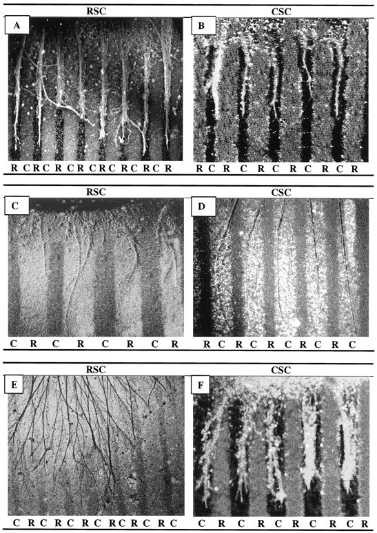
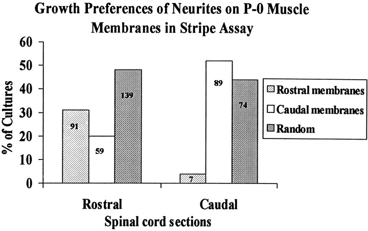
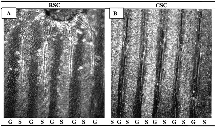
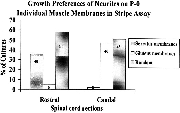
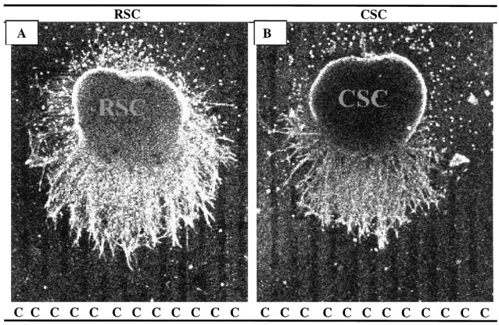
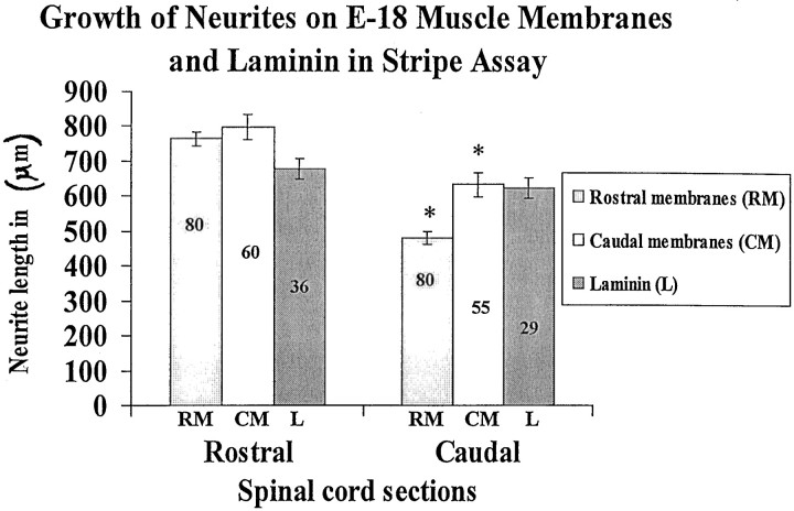
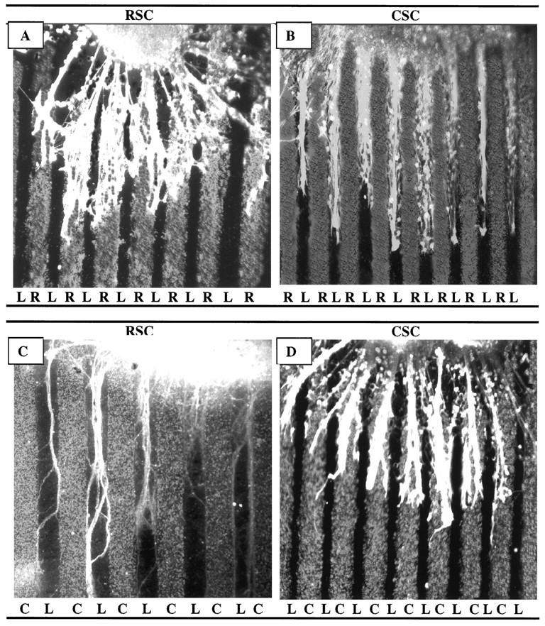
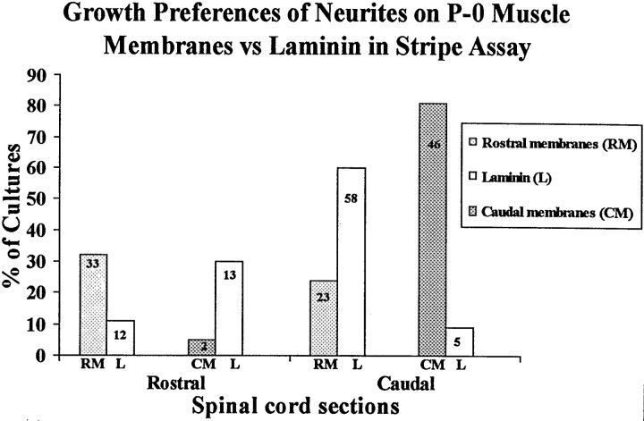
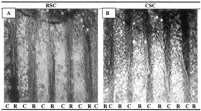
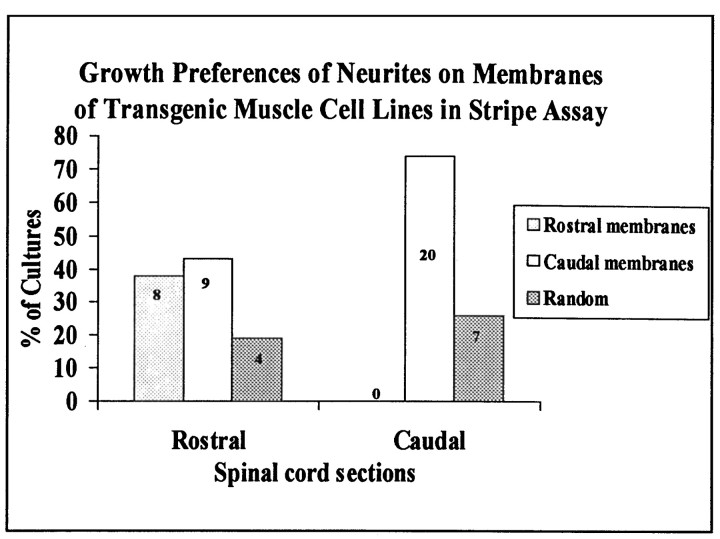
Motor neurons from distinct positions along the rostrocaudal axis generally innervate muscles or muscle fibers from corresponding axial levels. These topographic maps of connectivity are partially restored after denervation or transplantation under conditions in which factors of timing and proximity are eliminated. It is therefore likely that motor neurons and some intramuscular structures bear cues that bias synapse formation in favor of positionally matched partners. To localize these cues, we studied outgrowth of neurites from embryonic spinal cord explants on carpets of membranes isolated from perinatal rat muscles. Neurites from rostral (cervical) and caudal (lumbar) spinal cord slices exhibit distinct growth preferences. In many instances, rostrally derived neurites grew selectively on membranes from forelimb muscles or from a single thoracic muscle (the serratus anterior) when given a choice between these membranes and membranes from hindlimb muscles or laminin. Caudally derived neurites almost never exhibited such rostral preferences, but instead preferred membranes from hindlimb muscles or a single hindlimb muscle (the gluteus) to rostral muscles or laminin. Likewise, spinal neurites exhibited distinct position-related preferences for outgrowth on membranes of clonal myogenic cell lines derived from specific rostral and caudal muscles. Taken together these results suggest that the membranes of motor axons and myotubes bear complementary labels that vary with rostrocaudal position and regulate neuromuscular connectivity.


Figures

Fig. 1.
Fig. 1.
Position-dependent preferential outgrowth of spinal neurites on membranes from P0 and E18 limb muscles. Spinal cord slices cut from the cervical (rostral, RSC) or lumbar (caudal, CSC) enlargement were placed on alternating stripes of membrane derived from forelimb (rostral) or hindlimb (caudal) muscles. Membranes applied to one set of lanes in each culture were mixed with fluorescein-labeled beads to mark lane boundaries and marked rostral (R) or caudal (C). After 3 d, the cultures were fixed, and neurites were labeled with antibodies to neurofilaments (A, B), left unstained (C, D), or reacted with HRP-DAB(E) (see Materials and Methods). Rostral spinal cord neurites grew selectively on P0 rostral membranes in 31% of cultures(A) and on E18 rostral membranes in 9% of cultures(C). Caudal spinal cord neurites grew selectively on caudal P0 membranes in 52% of cultures (B) and on caudal E18 membranes in 78% of cultures (D). E, Rostral neurites grew randomly in 42% of cultures on E18 rostral membranes. F, Neurites of caudal spinal cord growing on caudal P0 membranes and stained with fluorescein-labeled antibody to acetylcholine transferase. Magnification: A, E, 60×;B, D, F, 100×; C, 140×.
Fig. 2.
Fig. 2.
Position-dependent preferential outgrowth of spinal neurites on membranes from P0 limb muscles. Cultures were prepared as in Figure 1. Columns show percentage of cultures showing preferential outgrowth on rostral or caudal membranes or no striking preference (Random). Numbers indicate number of cultures in each category.
Fig. 3.
Fig. 3.
Position-dependent preferential outgrowth of spinal neurites on membranes from individual muscles. Spinal cord slices cut from the cervical (rostral, RSC) or lumbar (caudal, CSC) enlargement were placed on alternating stripes of membrane derived from the serratus anterior (S), a rostral muscle, or from the gluteus (G), a caudal muscle. Membranes applied to one set of lanes in each culture were mixed with fluorescent beads to mark lane boundaries. After 3 d, the cultures were fixed, and neurites were labeled with antibodies to neurofilaments. (Figures shown are unstained.) A, Rostral spinal cord neurites grew selectively on serratus anterior membranes in 36% of cultures. B, Caudal neurites grew selectively on gluteus membranes in 47% of cultures. Magnification: A, 100×; B, 80×.
Fig. 4.
Fig. 4.
Position-dependent preferential outgrowth of spinal neurites on membranes from individual muscles. Cultures were prepared as in Figure 3. Columns show percentage of cultures showing preferential outgrowth on rostral (serratus anterior) or caudal (gluteus) membranes or no striking preference (Random).Numbers indicate number of cultures in each category.
Fig. 5.
Fig. 5.
Rostral spinal cord neurites grew longer than caudal neurites on membrane lanes. Spinal cord slices cut from the cervical (RSC) or lumbar (CSC) enlargement were placed on stripes of membrane derived from hindlimb (C, caudal) muscles (ventral surface of slice is down). Membranes mixed with fluorescent beads were applied to one set of lanes in each culture; intervening dark lanes were uncoated. After 3 d, the cultures were fixed, and neurites were labeled with antibodies to neurofilaments. A, Rostral neurites growing on caudal membranes. B, Caudal neurites growing on caudal membranes. Both A and B are at 40× magnification.
Fig. 6.
Fig. 6.
Spinal cord neurites grow similar lengths on rostral and caudal membranes and on laminin. Cultures were prepared as in Figure 5. Membranes or laminin were applied to one set of lanes in each culture. Intervening lanes were uncoated. Measurements of neurite length were made in cultures as in Figure 5. Columnsshow average neurite length (±SE), measured as the distance from the edge of the explant to the end of the longest neurite. Extent of outgrowth on laminin-coated stripes (data not shown, but see Figure 7) is given for comparison. Numbers indicate number of cultures in each category. Asterisks indicate that caudal neurites grew significantly less than rostral neurites on both rostral and caudal membranes (p < 0.05).
Fig. 7.
Fig. 7.
Spinal cord neurites make position-dependent choices between muscle membranes and laminin. Spinal cord slices cut from the cervical (rostral, RSC) or lumbar (caudal,CSC) enlargement were placed on alternating stripes of muscle membranes (R, rostral, or C, caudal) and laminin. A, Rostral spinal cord neurites prefer rostral membranes to laminin. B, Caudal neurites prefer laminin to rostral membranes. C, Rostral neurites prefer laminin to caudal membranes. D, Caudal neurites prefer caudal membranes to laminin. Neurites were stained with fluorescein–anti-NF. Magnification: A, 80×; B, D, 60×; C, 100×.
Fig. 8.
Fig. 8.
Spinal cord neurites make position-dependent choices between muscle membranes and laminin. Cultures were prepared as in Figure 7, in which alternating lanes contained rostral membranes, caudal membranes, or laminin. Columns show percentage of cultures showing preferential outgrowth on rostral or caudal membranes (RM or CM) or laminin (L).Numbers indicate number of cultures in each category. An additional 108 cultures, not included in this graph, did not show striking preferences for either membranes or laminin (59, 28, 15, and 6 in cocultures of the types shown in Figure 7A–D, respectively).
Fig. 9.
Fig. 9.
Position-dependent preferential outgrowth of spinal neurites on membranes from muscle cell lines. Spinal cord slices cut from the cervical (rostral, RSC) or lumbar (caudal,CSC) enlargement were placed on alternating stripes of myotube membranes prepared from immortalized cell lines. Membranes applied to one set of lanes in each culture were mixed with fluorescent beads to mark lane boundaries (R, rostral; C, caudal). After 3 d, the cultures were fixed, and neurites were labeled with fluorescein-labeled antibodies to neurofilaments.A, Rostral spinal cord neurites grew selectively on membranes from the rostrally derived cell lines. B, Caudal neurites grew selectively on membranes from the caudally derived cell lines. Magnification: 100×.
Fig. 10.
Fig. 10.
Position-dependent preferential outgrowth of spinal neurites on membranes from muscle cell lines. Cultures were prepared as in Figure 9. Columns show percentage of cultures showing preferential outgrowth on rostral or caudal membranes or no striking preference (Random). Numbers indicate number of cultures in each category.

Similar articles

Cited by

References

    1. Bennett MR, Lavidis NA. Segmental motor projections to rat muscles during the loss of polyneuronal innervation. Dev Brain Res. 1984a;13:1–7. - PubMed
    1. Bennett MR, Lavidis NA. Development of the topographical projection of motor neurons to a rat muscle accompanies loss of polyneuronal innervation. J Neurosci. 1984b;4:2204–2212. - PMC - PubMed
    1. Brown MC, Booth CM. Segregation of motor nerves on a segmental basis during synapse elimination in neonatal muscles. Brain Res. 1983a;273:188–190. - PubMed
    1. Brown MC, Booth CM. Postnatal development of the adult pattern of motor axon distribution in rat muscles. Nature. 1983b;304:741–742. - PubMed
    1. Brown MC, Hardman VJ. A reassessment of the accuracy of reinnervation by motoneurons following crushing or freezing of the sciatic or lumbar spinal nerves of rats. Brain. 1987;110:695–705. - PubMed

Publication types

MeSH terms

LinkOut - more resources