

Depression duration but not age predicts hippocampal volume loss in medically healthy women with recurrent major depression
- PMID: 10366636
- PMCID: PMC6782668
- DOI: 10.1523/JNEUROSCI.19-12-05034.1999
Depression duration but not age predicts hippocampal volume loss in medically healthy women with recurrent major depression
Abstract
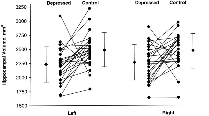
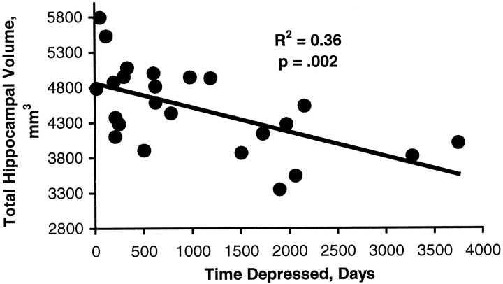
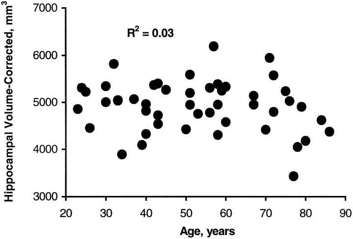
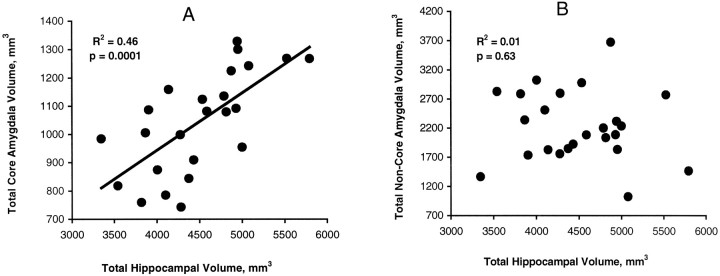
This study takes advantage of continuing advances in the precision of magnetic resonance imaging (MRI) to quantify hippocampal volumes in a series of human subjects with a history of depression compared with controls. We sought to test the hypothesis that both age and duration of past depression would be inversely and independently correlated with hippocampal volume. A sample of 24 women ranging in age from 23 to 86 years with a history of recurrent major depression, but no medical comorbidity, and 24 case-matched controls underwent MRI scanning. Subjects with a history of depression (post-depressed) had smaller hippocampal volumes bilaterally than controls. Post-depressives also had smaller amygdala core nuclei volumes, and these volumes correlated with hippocampal volumes. In addition, post-depressives scored lower in verbal memory, a neuropsychological measure of hippocampal function, suggesting that the volume loss was related to an aspect of cognitive functioning. In contrast, there was no difference in overall brain size or general intellectual performance. Contrary to our initial hypothesis, there was no significant correlation between hippocampal volume and age in either post-depressive or control subjects, whereas there was a significant correlation with total lifetime duration of depression. This suggests that repeated stress during recurrent depressive episodes may result in cumulative hippocampal injury as reflected in volume loss.
Figures
References
-
- Aboitz F, Scheibel A, Zaidel E. Morphometry of the Sylvian fissure and the corpus callosum, with emphasis on sex differences. Brain. 1992;115:1521–1541. - PubMed
-
- Amsterdam J, Maislin G, Berwish N, Phillips J, Winokur A. Enhanced adrenocortical sensitivity to submaximal doses of cosyntropin in depressed patients. Arch Gen Psychiatry. 1989;46:550–554. - PubMed
-
- Andreasen NC, Flashman L, Flaum M, Arndt S, Swayzee V, O’Leary DS, Ehrhardt JC, Yuh WTC. Regional brain abnormalities in schizophrenia measured with magnetic resonance imaging. JAMA. 1994;272:1763–1769. - PubMed
-
- Armanini MP, Hutchins C, Stein BA, Sapolsky RM. Glucocorticoid endangerment of hippocampal neurons is NMDA-receptor dependent. Brain Res. 1990;532:7–12. - PubMed
-
- Aronson S. J. Intracranial vascular lesions in patients with diabetes mellitus. Neuropathol Exp Neurol. 1973;32:183–196. - PubMed
Publication types
MeSH terms
Substances
Grants and funding
LinkOut - more resources
Full Text Sources
Other Literature Sources
Medical
Miscellaneous