. 1999 Jun;181(12):3632-43.
doi: 10.1128/JB.181.12.3632-3643.1999.

A Bacillus subtilis secreted protein with a role in endospore coat assembly and function

Affiliations

A Bacillus subtilis secreted protein with a role in endospore coat assembly and function

M Serrano et al. J Bacteriol. 1999 Jun.

Abstract

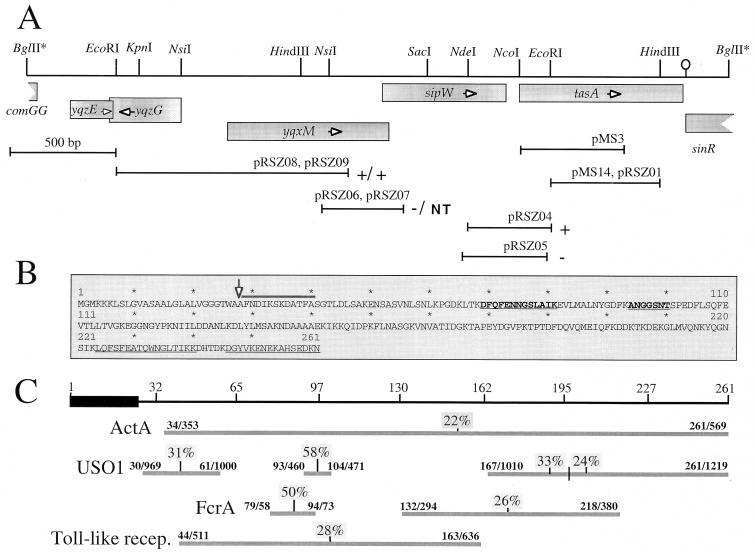
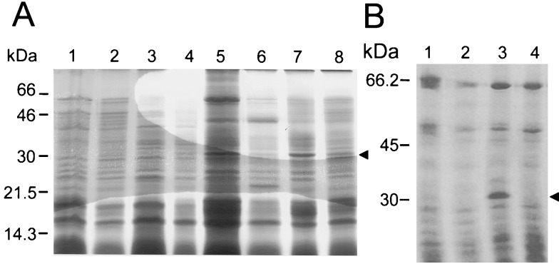
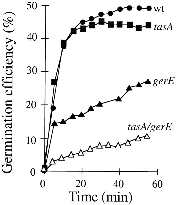
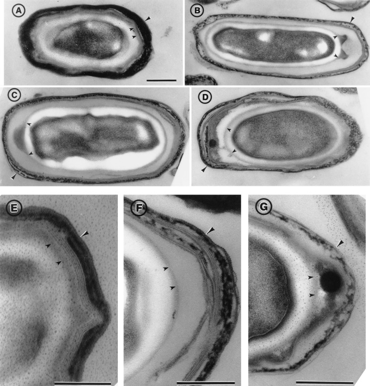
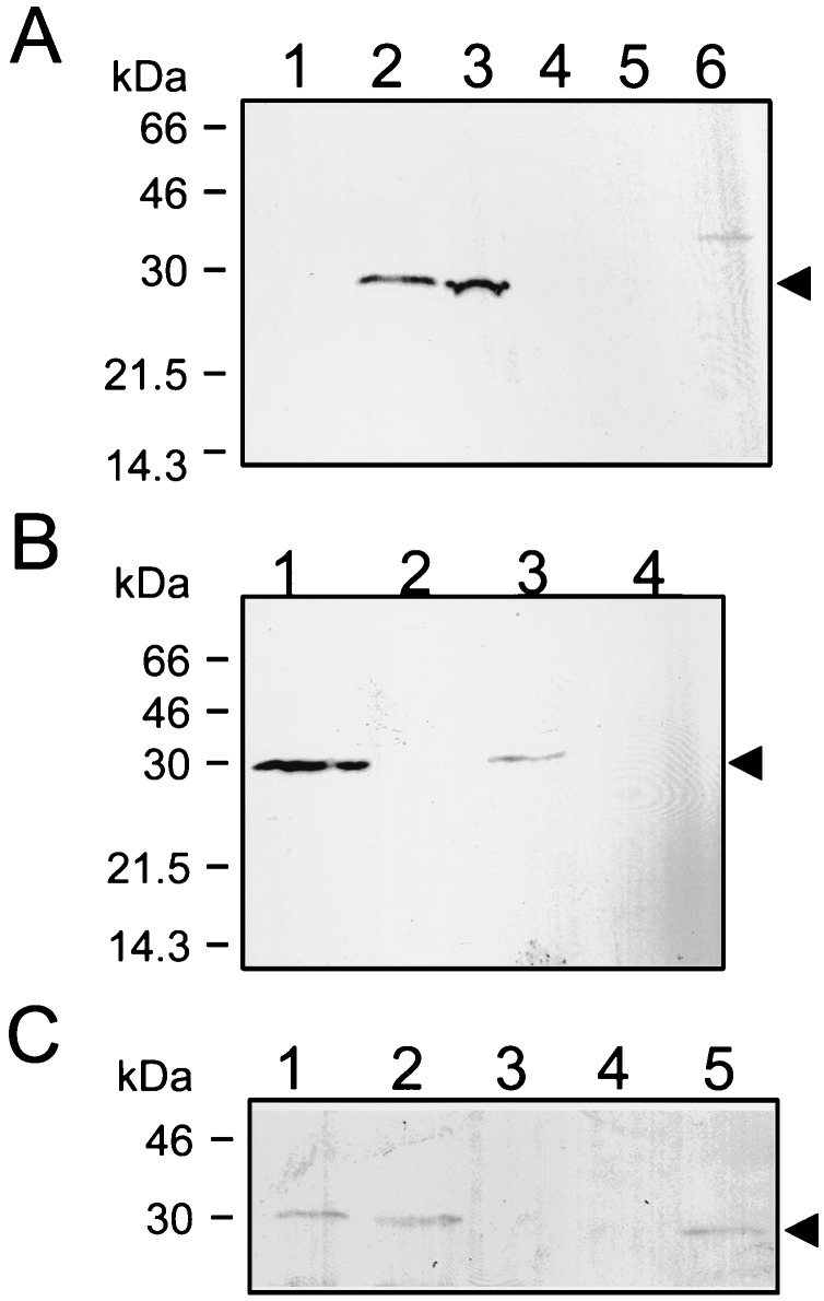
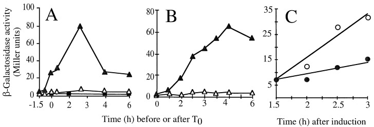
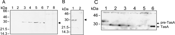
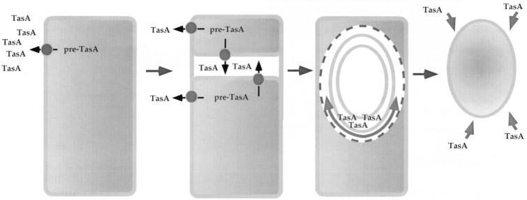
Bacterial endospores are encased in a complex protein coat, which confers protection against noxious chemicals and influences the germination response. In Bacillus subtilis, over 20 polypeptides are organized into an amorphous undercoat, a lamellar lightly staining inner structure, and an electron-dense outer coat. Here we report on the identification of a polypeptide of about 30 kDa required for proper coat assembly, which was extracted from spores of a gerE mutant. The N-terminal sequence of this polypeptide matched the deduced product of the tasA gene, after removal of a putative 27-residue signal peptide, and TasA was immunologically detected in material extracted from purified spores. Remarkably, deletion of tasA results in the production of asymmetric spores that accumulate misassembled material in one pole and have a greatly expanded undercoat and an altered outer coat structure. Moreover, we found that tasA and gerE mutations act synergistically to decrease the efficiency of spore germination. We show that tasA is the most distal member of a three-gene operon, which also encodes the type I signal peptidase SipW. Expression of the tasA operon is enhanced 2 h after the onset of sporulation, under the control of sigmaH. When tasA transcription is uncoupled from sipW expression, a presumptive TasA precursor accumulates, suggesting that its maturation depends on SipW. Mature TasA is found in supernatants of sporulating cultures and intracellularly from 2 h of sporulation onward. We suggest that, at an early stage of sporulation, TasA is secreted to the septal compartment. Later, after engulfment of the prespore by the mother cell, TasA acts from the septal-proximal pole of the spore membranes to nucleate the organization of the undercoat region. TasA is the first example of a polypeptide involved in coat assembly whose production is not mother cell specific but rather precedes its formation. Our results implicate secretion as a mechanism to target individual proteins to specific cellular locations during the assembly of the bacterial endospore coat.


Figures

FIG. 1
FIG. 1
Chromosomal organization and structure of tasA. (A) Partial restriction map and genetic organization of the tasA region. The boxes below the restriction map indicate the extent and direction of transcription of the different cistrons in the region, as deduced from the analysis of the B. subtilis genome sequence (22). The stem-and-loop structure downstream of tasA indicates the position of a possible transcription terminator. The lines below the restriction map represent DNA fragments cloned into the indicated plasmids. The plus or minus sign to the right of a plasmid denotes the Lac phenotype of a B. subtilis strain carrying a transcriptional fusion of the corresponding fragment to the lacZ gene inserted in single copy at the amyE locus or at the tasA region, respectively (see text). pRSZ04 (integrating at the tasA region) and pRSZ05 (amyE integrational) are shown in separate lines, since the inserts in each plasmid differ slightly. “NT” indicates that no transformants of pRSZ07 were obtained. (B) Deduced primary structure of TasA. The thick line above the sequence denotes the N-terminal amino acid sequence determined by the Edman reaction of the coat-associated TasA polypeptide. The first 23 residues of TasA are thought to be a signal peptide, and the processing site is indicated by a vertical arrow. The internal segments underlined were determined by MALDI mass spectrometry analysis, after cleavage of a six-His-S.Tag-TasA fusion protein with Lys-C protease (see Materials and Methods). The sequences in boldface were determined by the Edman reaction. (C) Regions of sequence similarity between TasA and the indicated proteins. The numbers above the horizontal lines indicate the residues that delimit those regions in both proteins. The numbers above the vertical lines indicate the percentages of sequence identity for the indicated segments. The ActA, USO1, FcrA, and Toll-like protein sequences have the following database accession numbers: S20887, Z74106, S35760, and U88879, respectively. recep., receptor.
FIG. 2
FIG. 2
Extraction of TasA from gerE mutant spores. Spores of a wild-type strain and various mutant strains were purified, and the coat proteins were extracted by treatment with a buffer containing SDS-DTT (A) or with alkali (B). The extracted proteins were resolved in 12.5% polyacrylamide gels containing SDS, and the gels were stained with Coomassie brilliant blue. The spores used for the extraction of the coat proteins were as follows: (A) lane 1, wild type; lane 2, tasA mutant; lane 3, cotE mutant; lane 4, cotE tasA mutant; lane 5, gerE mutant; lane 6, gerE tasA mutant; lane 7, cotE gerE mutant; lane 8, gerE cotE tasA mutant; (B) lane 1, wild type; 2, tasA mutant; lane 3, gerE mutant; lane 4, gerE tasA mutant. The arrowheads indicate the position of the 30-kDa TasA polypeptide which is readily extracted from gerE mutant spores. Also indicated are the positions of the molecular mass markers (in kilodaltons).
FIG. 3
FIG. 3
Germination efficiency of wild-type and mutant spores. Spores produced by a wild type (wt), as well as by tasA, gerE, and gerE tasA mutants, were purified, and the kinetics of germination were examined. Germination was induced by l-alanine in GFK, as previously described (27), and monitored by the decrease in OD600 of the spore suspension. The efficiency of germination is defined as the ratio between the optical density of the culture at a given time after exposure to the germinant mixture and the original optical density of the suspension.
FIG. 4
FIG. 4
Electron microscopy of wild-type and tasA mutant spores. Spores were collected from DSM cultures of a wild type (MB24 [A and E]) and the tasA mutant (AH1700 [B to D, F, and G]), 48 h after the initiation of sporulation. The spores were purified by centrifugation through Renocal gradients and processed for electron microscopy analysis as described in Materials and Methods. The large arrowheads point to the outer coat structure. The smaller arrowheads indicate the boundary between the cortex and the undercoat, which is probably defined by the forespore outer membrane. Darkly staining material (between the two small arrowheads) accumulates in the undercoat region of the tasA mutant but not in wild-type spores. Scale bar, 0.2 μm.
FIG. 5
FIG. 5
Detection of TasA in wild-type and mutant spores. Immunoblot analysis of material extracted from purified spores produced by a wild-type strain and various mutant strains. (A) Spores of a wild-type strain (MB24, lane 1) or of the following mutant strains were purified 24 h after the onset of sporulation: cotE mutant (AH1823, lane 2), gerE mutant (AH94, lane 3), tasA mutant (AH1822, lane 4), cotE tasA mutant (AH1824, lane 5), and gerE tasA mutant (AH1827, lane 6). (B) Spores used were purified 48 h after the onset of sporulation from cultures of a wild-type strain (MB24, lane 1), a tasA mutant (AH1822, lane 2), a gerE mutant (AH94, lane 3), or a gerE tasA double mutant (AH1827, lane 4). (C) Coat proteins were extracted from purified spores before or after washing of the spore suspension with 1 M KCl (45). Lane 1, wild type, washed; lane 2, wild type, before washing; lane 3, tasA mutant, not washed; lane 4, gerE mutant, washed; lane 5, gerE mutant, before washing. Proteins were resolved on SDS-containing 12.5% polyacrylamide gels and transferred to nitrocellulose membranes. The membranes were then probed with an anti-TasA antiserum. The arrowheads indicate the positions of TasA antigen. The positions of molecular mass markers (in kilodaltons) are also indicated.
FIG. 6
FIG. 6
tasA is controlled by ςH. The figure illustrates the time course of β-galactosidase production by various strains bearing a tasA-lacZ transcriptional fusion integrated at the tasA locus. Different growth conditions were used. (A) Strains were induced to sporulate in DSM, and T0 defines the onset of sporulation. (B) YT medium (2×) was used (T0 corresponds to an OD600 of about 0.3, whereas maximum enzyme activity was reached about 3 to 4 h later). Enzyme production was measured in strain AZ404 (tasA-lacZ) (dark triangles) and its congenic sigH mutant (AZ393, dark squares). (C) A Pspac-spo0H strain carrying the tasA-lacZ fusion was grown in 2× YT medium to a low OD600 value (about 0.2), at which point the culture was divided in half. IPTG was added to one flask (open circles) but not to the other (closed circles). Samples were collected at the indicated times, and the specific activity of β-galactosidase was determined with the substrate o-nitrophenol-β-d-galactopyranoside (ONPG). Background levels of β-galactosidase synthesis were estimated for the wild-type strain PY79 (open triangles in panels A and B).
FIG. 7
FIG. 7
TasA production and pre-TasA processing during growth and sporulation. (A) Samples of DSM cultures of strain AH94 (gerE36) were harvested during the logarithmic phase of growth (lane 1), at T0 (lane 2), T2 (lane 3), T4 (lane 4), and T6 (lane 5), and whole-cell extracts were prepared. The cultures were allowed to sporulate, and coat material was then isolated from spores purified 24 h after T0 (lane 6). DSM cultures of a ςH mutant (strain AH17; lane 7) or of a gerE tasA insertional mutant (strain AH1827, lane 8) were also collected at T2. (B) Samples of supernatants of a culture of a tasA+ strain (AH94, lane 1) or of a tasA mutant (AH1700, lane 2) were collected at T2, and the proteins were concentrated as described in Materials and Methods. (C) The inducer IPTG was added to a DSM culture of the Pspac-tasA strain AH1802 at T0. Samples were collected at 30 (lane 1), 60 (lane 2), 90 (lane 3), and 120 min (lane 4) after the addition of IPTG, and whole-cell lysates were prepared. Samples of DSM cultures of a tasA mutant (lane 5) and of a tasA+ strain (lane 6) at T2 were also analyzed. Proteins in all samples were resolved on 12.5% polyacrylamide gels containing SDS and transferred to nitrocellulose membranes. The membranes were probed with an anti-TasA antiserum. The arrows indicate the positions of the TasA antigen. Molecular mass markers (in kilodaltons) are also indicated.
FIG. 8
FIG. 8
Model for TasA function in coat assembly. The figure illustrates the various morphological stages of development in relation to TasA production. TasA production is initiated under ςH command early in sporulation, before the asymmetric division that marks morphological stage II. In this early period, TasA is secreted to the culture medium, where its mature form accumulates. Processing is thought to rely on the SipW type I signal peptidase. After formation of the sporulation septum, the mature form of TasA accumulates in the space defined by the two septal membranes. After engulfment, TasA may localize preferentially on the septum-proximal side of the spore. From this position, TasA may nucleate the organization of the undercoat material, which then proceeds around the entire forespore. In the absence of TasA, a scar is left at the nucleation position. Finally, after release of the spore upon lysis of the mother cell, TasA in the culture medium can associate with the spore coats. Alternatively (not represented), in old spores, the spore-associated TasA becomes more extractable.

Similar articles

Cited by

References

    1. Aronson A I, Fitz-James P. Structure and morphogenesis of the bacterial spore coat. Bacteriol Rev. 1976;40:360–402. - PMC - PubMed
    1. Aronson A I, Song H-Y, Bourne N. Gene structure and precursor processing of a novel Bacillus subtilisspore coat protein. Mol Microbiol. 1988;3:437–444. - PubMed
    1. Aronson A I, Ekanayake L, Fitz-James P C. Protein filaments may initiate the assembly of the Bacillus subtilisspore coat. Biochimie. 1992;74:661–667. - PubMed
    1. Beall B, Driks A, Losick R, Moran C P., Jr Cloning and characterization of a gene required for assembly of the Bacillus subtilisspore coat. J Bacteriol. 1993;175:1705–1716. - PMC - PubMed
    1. Benson A K, Haldenwang W G. Regulation of ςB levels and activity in Bacillus subtilis. J Bacteriol. 1993;175:2347–2356. - PMC - PubMed

Publication types

LinkOut - more resources