. 1999 Jun;181(12):3710-5.
doi: 10.1128/JB.181.12.3710-3715.1999.

The morphological transition of Helicobacter pylori cells from spiral to coccoid is preceded by a substantial modification of the cell wall

Affiliations

The morphological transition of Helicobacter pylori cells from spiral to coccoid is preceded by a substantial modification of the cell wall

K Costa et al. J Bacteriol. 1999 Jun.

Abstract

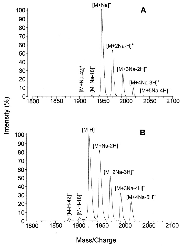
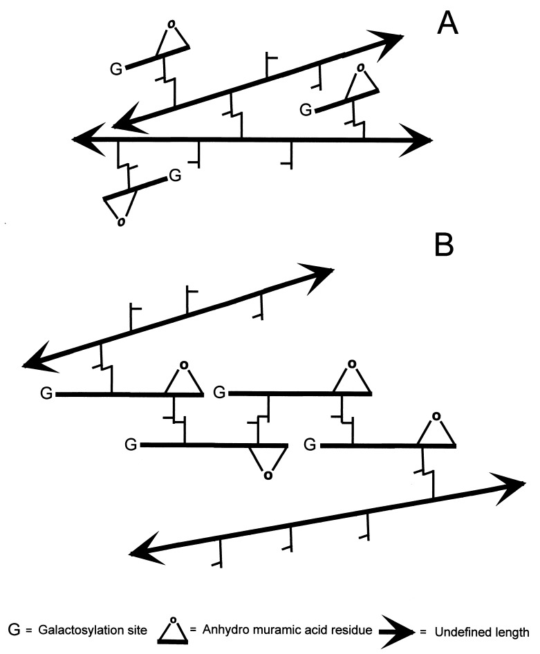
The peptidoglycan (murein) of Helicobacter pylori has been investigated by high-performance liquid chromatography and mass spectrometric techniques. Murein from H. pylori corresponded to the A1gamma chemotype, but the muropeptide elution patterns were substantially different from the one for Escherichia coli in that the former produced high proportions of muropeptides with a pentapeptide side chain (about 60 mol%), with Gly residues as the C-terminal amino acid (5 to 10 mol%), and with (1-->6)anhydro-N-acetylmuramic acid (13 to 18 mol%). H. pylori murein also lacks murein-bound lipoprotein, trimeric muropeptides, and (L-D) cross-linked muropeptides. Cessation of growth and transition to coccoid shape triggered an increase in N-acetylglucosaminyl-N-acetylmuramyl-L-Ala-D-Glu (approximately 20 mol%), apparently at the expense of monomeric muropeptides with tri- and tetrapeptide side chains. Muropeptides with (1-->6)anhydro-muramic acid and with Gly were also more abundant in resting cells.


Figures

FIG. 1
FIG. 1
HPLC elution patterns of murein samples purified from spiral and coccoid H. pylori cells. Muropeptide mixtures were analyzed as described in the text, and the A204 of the eluent was monitored. A murein sample from E. coli was analyzed under identical conditions for comparative purposes. Numbers in H. pylori panels identify corresponding peaks in both spiral and coccoid cell samples. Muropeptides shown in the E. coli panel correspond to the basic structure N-acetylglucosaminyl-N-acetylmuramyl–l-Ala–d-Glu–(γ)-mDap–R1R2, where R1 and R2 are substituents at the l-carboxy and d-amino groups of mDap, respectively. R1 and R2 for the muropeptides shown are as follows: A, R1 = →d-Ala, R2 = −H; B, R1 = →Lys-Arg, R2 = −H (Braun’s lipoprotein anchoring muropeptide); C, R1 = N-acetylglucosaminyl-N-acetylmuramyl–l-Ala–d-Glu–(d-Ala)mDap←, R2 = −H; D, R1 = N-acetylglucosaminyl-N-acetylmuramyl–l-Ala–d-Glu–mDap–d-Ala←, R2 = −H; E, R1 = N-acetylglucosaminyl-N-acetylmuramyl–l-Ala–d-Glu–(d-Ala)mDap–d-Ala←, R2 = −H; F, R1 = N - acetylglucosaminyl - N - acetylmuramyl–l - Ala–d - Glu–(d - Ala)mDap–d - Ala ←, R2 = N-acetylglucosaminyl-N-acetylmuramyl–l-Ala–d-Glu–mDap–d-Ala→.
FIG. 2
FIG. 2
Molecular ion region of the positive- and negative-ion MALDI mass spectrum of fraction 7 from murein of spiral H. pylori cells. (A) The positive-ion mass spectrum exhibited complex adduct ions at m/z 1945.0 (calculated m/z, 1945.9), 1966.9, 1989.0, 2011.1, and 2033.0, corresponding to [M+Na]+, [M+2Na-H]+, [M+3Na-2H]+, [M+4Na-3H]+, and [M+5Na-4H]+ ions, respectively. (B) In the negative-ion mass spectrum, a similar complex adduct ion pattern ([M+Na-2H], [M+2Na-3H], [M+3Na-4H], and [M+4Na-5H]) was observed, but with the deprotonated molecule [M-H] (found m/z, 1921.0; calculated, m/z 1921.9) as the base peak.
FIG. 3
FIG. 3
Alternative models accounting for the high proportion in cross-linked anhydro-muropeptides susceptible to galactosylation. (A) Single anhydro-disaccharide units are cross-linked to nearby long glycan strands. Muropeptides with this configuration do not contribute to the physical strength of the sacculus. (B) Short strands (two disaccharide units) are head-to-tail cross-linked to each other, connecting longer strands. In this configuration very short chains could effectively contribute to the strength of the sacculus. Muramidase digestion would release equal amounts of galactosylated, anhydro-cross-linked dimers in both instances. The models shown in both panels could coexist.

Similar articles

Cited by

References

    1. Aleljung P, Nilsson H O, Wang X, Nyberg P, Morner T, Warsame I, Wadstrom T. Gastrointestinal colonisation of BALB/cA mice by Helicobacter pylorimonitored by heparin magnetic separation. FEMS Immunol Med Microbiol. 1996;13:303–309. - PubMed
    1. Benaïssa M, Babin P, Quellard N, Pezennec L, Cenatiempo Y, Fauchère J L. Changes in Helicobacter pyloriultrastructure and antigens during conversion from the bacillary to the coccoid form. Infect Immun. 1996;64:2331–2335. - PMC - PubMed
    1. Blaser M J. Helicobacter pyloriand the pathogenesis of gastroduodenal inflammation. J Infect Dis. 1990;161:626–633. - PubMed
    1. Bode G, Mauch F, Malfertheiner P. The coccoid forms of Helicobacter pylori. Criteria for their viability. Epidemiol Infect. 1993;111:483–490. - PMC - PubMed
    1. Braun V, Wu H C. Lipoproteins, structure, function, biosynthesis and model for protein export. In: Ghuysen J M, Hakenbeck R, editors. Bacterial cell wall. Amsterdam, The Netherlands: Elsevier Science Publisher; 1994. pp. 319–341.

Publication types

MeSH terms