. 1999 Jun;181(12):3730-42.
doi: 10.1128/JB.181.12.3730-3742.1999.

Bacterioferritin A modulates catalase A (KatA) activity and resistance to hydrogen peroxide in Pseudomonas aeruginosa

Affiliations

Bacterioferritin A modulates catalase A (KatA) activity and resistance to hydrogen peroxide in Pseudomonas aeruginosa

J F Ma et al. J Bacteriol. 1999 Jun.

Abstract

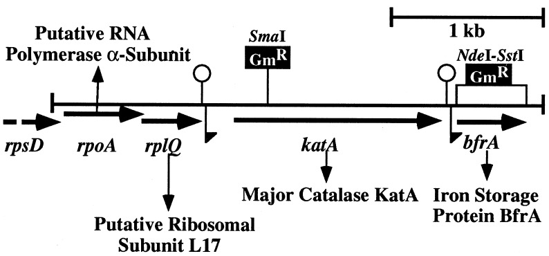
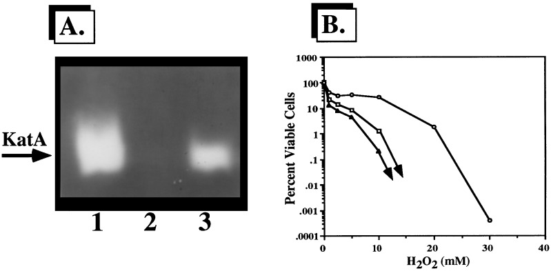
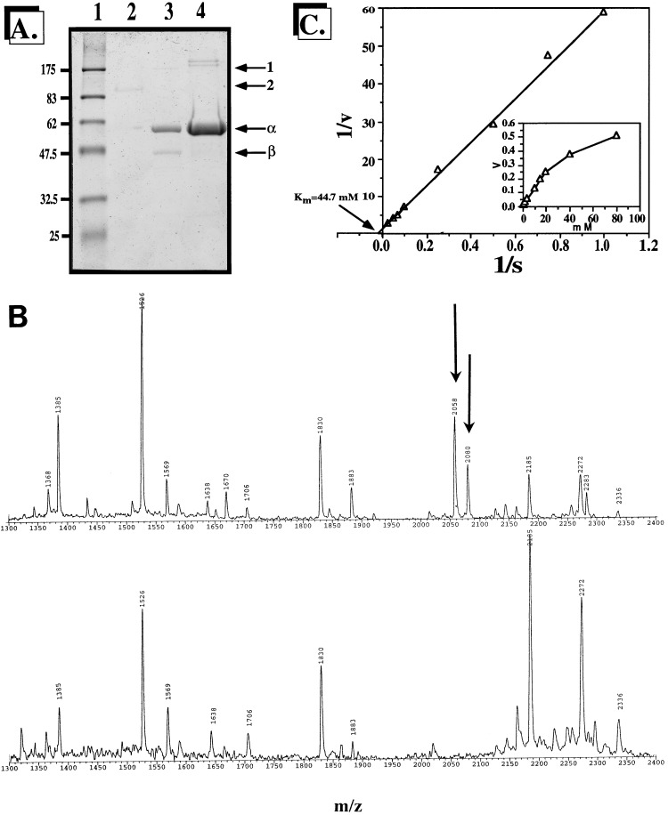
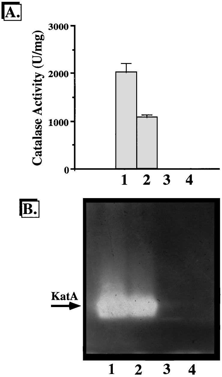
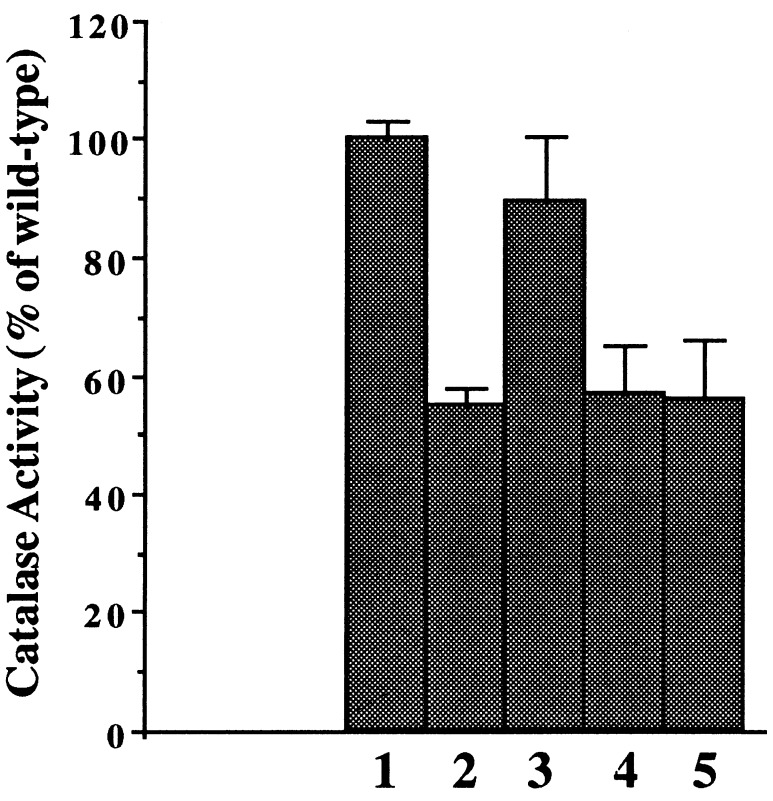
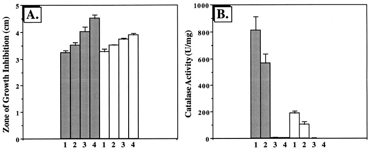
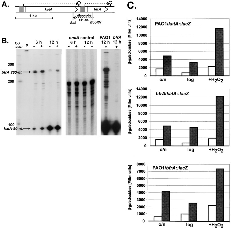
We have cloned a 3.6-kb genomic DNA fragment from Pseudomonas aeruginosa harboring the rpoA, rplQ, katA, and bfrA genes. These loci are predicted to encode, respectively, (i) the alpha subunit of RNA polymerase; (ii) the L17 ribosomal protein; (iii) the major catalase, KatA; and (iv) one of two iron storage proteins called bacterioferritin A (BfrA; cytochrome b1 or b557). Our goal was to determine the contributions of KatA and BfrA to the resistance of P. aeruginosa to hydrogen peroxide (H2O2). When provided on a multicopy plasmid, the P. aeruginosa katA gene complemented a catalase-deficient strain of Escherichia coli. The katA gene was found to contain two translational start codons encoding a heteromultimer of approximately 160 to 170 kDa and having an apparent Km for H2O2 of 44.7 mM. Isogenic katA and bfrA mutants were hypersusceptible to H2O2, while a katA bfrA double mutant demonstrated the greatest sensitivity. The katA and katA bfrA mutants possessed no detectable catalase activity. Interestingly, a bfrA mutant expressed only approximately 47% the KatA activity of wild-type organisms, despite possessing wild-type katA transcription and translation. Plasmids harboring bfrA genes encoding BfrA altered at critical amino acids essential for ferroxidase activity could not restore wild-type catalase activity in the bfrA mutant. RNase protection assays revealed that katA and bfrA are on different transcripts, the levels of which are increased by both iron and H2O2. Mass spectrometry analysis of whole cells revealed no significant difference in total cellular iron levels in the bfrA, katA, and katA bfrA mutants relative to wild-type bacteria. Our results suggest that P. aeruginosa BfrA may be required as one source of iron for the heme prosthetic group of KatA and thus for protection against H2O2.


Figures

FIG. 1
FIG. 1
Genetic map of the 3.605-kb EcoRI-EcoRV fragment harboring rpoA, rplQ, katA, and bfrA of P. aeruginosa PAO1 and putative gene products. The large loopholes indicate transcriptional terminators. The flags pointing downward indicate the promoter regions upstream of the katA and bfrA genes. The restriction sites from which single katA and bfrA mutants were generated via insertional mutagenesis are given.
FIG. 2
FIG. 2
Amino acid similarity of BfrA and other Bfr proteins. (A) Proteins were aligned with Align Plus 3.0. Dots indicate identical amino acids, while dashes indicate gaps in the protein sequence relative to P. aeruginosa BfrA. PA, P. aeruginosa BfrA; PP, P. putida Bfr; NG, N. gonorrhoeae Bfr; ST, S. typhimurium Bfr; EC, E. coli Bfr. The amino acids essential for ferroxidase activity in E. coli Bfr are conserved in each Bfr protein and are shown in boldface and marked with an asterisk. (B) Unrooted phylogenetic tree based on the amino acid sequences of 35 (bacterio)ferritins and 5 rubrerythrins and constructed by parsimony methods. The branch lengths (shown in italics) reflect the evolutionary distances calculated as the average number of amino acid changes per 1,000 residues. The numbers in bold in front of the nodes represent the proportion of bootstrap samplings that support the topology shown. Two hundred bootstrap replicates were analyzed. For analysis of the BfrA protein, Bfr sequences were obtained from GenBank (accession no.) for the following organisms: Arcfu, Archaeoglobus fulgidus putative ferritin (AE001047); Arcfur, A. fulgidus rubrerythrin 1 (AE001047); Azovi, Azotobacter vinelandii Bfr (U83692); Brume, Brucella melitensis Bfr (U19760); Caeel, Caenorhabditis elegans ferritin (AF106592); Camje, Campylobacter jejuni ferritin (D64082); Cloper, Clostridium perfringens rubreythrin (X92844); Desvur, Desulfovibrio vulgaris rubrerythrin (U82323); Echgr, Echinococcus granulosus ferritin (Z31712); Ecoli1, Escherichia coli ECOR30 Bfr (AF058450); Ecoli2, E. coli K-12 Bfr (M27176); Ecoli3, E. coli K-12 MG1655 cytoplasmic ferritin (AF000335); Galga, Gallus gallus ferritin heavy (H) chain (Y14698); Helpy, Helicobacter pylori J99 non-heme iron-containing ferritin Pfr (AE00149); Homsa, Homo sapiens apoferritin H chain (X00318); Ixori, Ixodes ricinus ferritin (AF068224); Lymst, Lymnaea stagnalis snail soma ferritin (P42577/X56778); Magma1, Magnetospirillum magnetotacticum Bfr1 (AF001959); Magma2, M. magnetotacticum Bfr2 (AF001959); Metth1, Methanobacterium thermoautotrophicum putative ferritin (AE000804); Metthr, M. thermoautotrophicum rubrerythrin (AE000854); Matjar, Methanococcus jannaschii rubrerythrin (U67520); Mycav, Mycobacterium avium Bfr (X76906); Mycle, Mycobacterium leprae Bfr (P43315); Myctu, Mycobacterium tuberculosis Bfr (Z97193); Neigo, Neisseria gonorrhoeae Bfr (U76633); Oncmy, Oncorhynchus mykiss ferritin-1 H chain (D86625); Ornmo, Ornithodoros moubata ferritin (AF068225); Porgi, Porphyromonas gingivalis ferritin (AB016086); Psepu: Pseudomonas putida Bfr (U66717); Pseae: Pseudomonas aeruginosa Bfr (AF047025); Ranca, Rana catesbeiana ferritin, middle subunit (J02724); Ratno, Rattus norvegicus ferritin H chain (P19132); Rhoca, Rhodobacter capsulatus Bfr (Z54247); Salty, Salmonella typhimurium LT2 Bfr (AF058449); SchjaH, Schistosoma japonicum putative ferritin-1 H chain (AF040385); SchmaL, Schistosoma mansoni ferritin light chain (M64538); Serma, Serratia marcescens Bfr (AF058451); Synec, Synechocystis sp. strain PCC6803 Bfr (D90905); and Wolba, Wolbachia sp. Bfr (21).
FIG. 2
FIG. 2
Amino acid similarity of BfrA and other Bfr proteins. (A) Proteins were aligned with Align Plus 3.0. Dots indicate identical amino acids, while dashes indicate gaps in the protein sequence relative to P. aeruginosa BfrA. PA, P. aeruginosa BfrA; PP, P. putida Bfr; NG, N. gonorrhoeae Bfr; ST, S. typhimurium Bfr; EC, E. coli Bfr. The amino acids essential for ferroxidase activity in E. coli Bfr are conserved in each Bfr protein and are shown in boldface and marked with an asterisk. (B) Unrooted phylogenetic tree based on the amino acid sequences of 35 (bacterio)ferritins and 5 rubrerythrins and constructed by parsimony methods. The branch lengths (shown in italics) reflect the evolutionary distances calculated as the average number of amino acid changes per 1,000 residues. The numbers in bold in front of the nodes represent the proportion of bootstrap samplings that support the topology shown. Two hundred bootstrap replicates were analyzed. For analysis of the BfrA protein, Bfr sequences were obtained from GenBank (accession no.) for the following organisms: Arcfu, Archaeoglobus fulgidus putative ferritin (AE001047); Arcfur, A. fulgidus rubrerythrin 1 (AE001047); Azovi, Azotobacter vinelandii Bfr (U83692); Brume, Brucella melitensis Bfr (U19760); Caeel, Caenorhabditis elegans ferritin (AF106592); Camje, Campylobacter jejuni ferritin (D64082); Cloper, Clostridium perfringens rubreythrin (X92844); Desvur, Desulfovibrio vulgaris rubrerythrin (U82323); Echgr, Echinococcus granulosus ferritin (Z31712); Ecoli1, Escherichia coli ECOR30 Bfr (AF058450); Ecoli2, E. coli K-12 Bfr (M27176); Ecoli3, E. coli K-12 MG1655 cytoplasmic ferritin (AF000335); Galga, Gallus gallus ferritin heavy (H) chain (Y14698); Helpy, Helicobacter pylori J99 non-heme iron-containing ferritin Pfr (AE00149); Homsa, Homo sapiens apoferritin H chain (X00318); Ixori, Ixodes ricinus ferritin (AF068224); Lymst, Lymnaea stagnalis snail soma ferritin (P42577/X56778); Magma1, Magnetospirillum magnetotacticum Bfr1 (AF001959); Magma2, M. magnetotacticum Bfr2 (AF001959); Metth1, Methanobacterium thermoautotrophicum putative ferritin (AE000804); Metthr, M. thermoautotrophicum rubrerythrin (AE000854); Matjar, Methanococcus jannaschii rubrerythrin (U67520); Mycav, Mycobacterium avium Bfr (X76906); Mycle, Mycobacterium leprae Bfr (P43315); Myctu, Mycobacterium tuberculosis Bfr (Z97193); Neigo, Neisseria gonorrhoeae Bfr (U76633); Oncmy, Oncorhynchus mykiss ferritin-1 H chain (D86625); Ornmo, Ornithodoros moubata ferritin (AF068225); Porgi, Porphyromonas gingivalis ferritin (AB016086); Psepu: Pseudomonas putida Bfr (U66717); Pseae: Pseudomonas aeruginosa Bfr (AF047025); Ranca, Rana catesbeiana ferritin, middle subunit (J02724); Ratno, Rattus norvegicus ferritin H chain (P19132); Rhoca, Rhodobacter capsulatus Bfr (Z54247); Salty, Salmonella typhimurium LT2 Bfr (AF058449); SchjaH, Schistosoma japonicum putative ferritin-1 H chain (AF040385); SchmaL, Schistosoma mansoni ferritin light chain (M64538); Serma, Serratia marcescens Bfr (AF058451); Synec, Synechocystis sp. strain PCC6803 Bfr (D90905); and Wolba, Wolbachia sp. Bfr (21).
FIG. 3
FIG. 3
(A) Complementation of P. aeruginosa katA in E. coli UM1. Cell extracts (20 μg) of aerobically grown, stationary-phase organisms were separated by nondenaturing PAGE and stained for catalase activity (51). Lane 1, P. aeruginosa PAO1; lane 2, E. coli UM1; lane 3, E. coli UM1(pJFM12). (B) Enhanced resistance of E. coli UM1 harboring P. aeruginosa katA to H2O2. Mid-logarithmic-phase bacteria were exposed to various concentrations of H2O2 for 15 min at 37°C (23). The results are typical of three separate experiments. Symbols: □, E. coli CSH7; ▴, E. coli UM1; ○, E. coli UM1(pJFM12).
FIG. 4
FIG. 4
Purification (A), mass spectrometric analysis (B), and Km measurement (C) of P. aeruginosa KatA. (A) Lane 1, protein molecular mass standards; lane 2, 60 ng of purified unboiled KatA; lane 3, 120 ng of purified boiled KatA; lane 4, 160 ng of purified boiled recombinant six-His-tagged KatA. (B) Mass spectrometric analysis of the 55-kDa subunit (top panel) and smaller subunits (bottom panel) of KatA. The arrows in the top panel indicate peptides that are absent in the bottom panel. (C) Double-reciprocal Lineweaver-Burk plot of KatA activity with various H2O2 concentrations. Experiments were performed as described in Materials and Methods.
FIG. 5
FIG. 5
Catalase activity (A) and activity staining (B) of P. aeruginosa strains. (A) Catalase activity in cell extracts from stationary-phase organisms was measured as described by Beers and Sizer (5); the values are means ± standard errors for three experiments. 1, P. aeruginosa PAO1; 2, bfrA; 3, katA; 4, katA bfrA. (B) Cell extracts (20 μg) from the above organisms were separated by nondenaturing PAGE in triplicate and stained for catalase activity (51). Lane 1, P. aeruginosa PAO1; lane 2, bfrA; lane 3, katA; lane 4, katA bfrA. The KatA activity band is shown by an arrow.
FIG. 6
FIG. 6
Importance of ferroxidase-center amino acids in optimal KatA activity in P. aeruginosa. P. aeruginosa harboring plasmids with wild-type or altered bfrA genes was grown aerobically to the stationary phase in L broth at 37°C. Catalase activity of cell extracts was monitored in triplicate. 1, Wild type plus pUCP19) 2, bfrA plus pUCP19) 3, bfrA plus pBFR4; 4, bfrA plus pBFR18 (E18K); 5, bfrA plus pBFR25 (Y25I).
FIG. 7
FIG. 7
H2O2 disk sensitivity (A) and catalase activity (B) of P. aeruginosa strains: influence of iron. (A) P. aeruginosa strains were grown aerobically in M9 minimal medium with or without 50 μM FeCl3 to the stationary phase. Organisms were diluted 30-fold in 3 ml of molten 0.6% M9 top agar and layered on M9 agar plates. Sterile filter paper disks (7 mm) were impregnated with 10 μl of 8.8 M H2O2 and placed in triplicate on the top-agar surface, and the plates were incubated at 37°C for 17 h. Zones of growth inhibition were measured. Shaded columns, M9 medium plus 50 μM FeCl3; open columns, M9 medium alone. The results are expressed as the means ± standard errors for nine different experiments. 1, P. aeruginosa PAO1; 2, bfrA; 3, katA; 4, katA bfrA. (B) Catalase activity (5) was measured in cell extracts from each strain and expressed as the means ± standard errors for three different experiments. Columns are as in panel A.
FIG. 8
FIG. 8
Regulation of katA and bfrA: effect of growth phase, iron, and H2O2. (A) Genetic organization of katA and bfrA and location of the riboprobe used for the simultaneous detection of the corresponding transcripts. The promoter regions upstream of katA and bfrA are depicted as shaded boxes. Loopholes indicate transcriptional terminators. nt., nucleotides. (B) RNase protection analysis of katA and bfrA. P. aeruginosa was grown aerobically under low (−)- or high (+)-iron conditions. An RNA ladder, undigested probe (P), and detected transcripts for bfrA and katA are indicated (left panel), together with omlA as a constitutive and loading control (middle panel) and the katA transcript in the wild type and the bfrA mutant (right panel). (C) Translational katA-lacZ and bfrA-lacZ activities. All bacteria were grown aerobically in M9 medium. Open columns, M9 low-iron medium (0.2 mM dipyridyl); shaded columns, M9 high-iron medium (50 μg of FeCl3 per ml). The three data pairs in each panel reflect the measured activities during the stationary phase after overnight growth (o/n), during exponential growth (log), and after 1 h of treatment with 1 mM H2O2 every 10 min (+H2O2). The values are the averages for quadruplicate cultures, and error bars (<10%) are omitted for clarity.

Similar articles

Cited by

References

    1. Altschul S F, Gish W, Miller W, Myers E W, Lipman D J. Basic local alignment search tool. J Mol Biol. 1990;215:403–410. - PubMed
    1. Andrews S C, Smith J M, Yewdall S J, Guest J R, Harrison P M. Bacterioferritins and ferritins are distantly related in evolution: conservation of ferroxidase-centre residues. FEBS Lett. 1991;293:164–168. - PubMed
    1. Ausubel F M, Brent R, Kingston R E, Moore D D, Seidman J D, Struhl K. Current protocols in molecular biology. New York, N.Y: John Wiley & Sons, Inc.; 1993. pp. 5.3.2–5.3.8.
    1. Bando Y, Aki K. Superoxide-mediated release of iron from ferritin by some flavoenzymes. Biochem Biophys Res Commun. 1990;168:389–395. - PubMed
    1. Beers R F, Jr, Sizer I W. A spectrophotometric method for measuring the breakdown of hydrogen peroxide by catalase. J Biol Chem. 1952;195:133–140. - PubMed

Publication types

MeSH terms

Associated data

LinkOut - more resources