

Case Reports
Treatment of distal aneurysms of the cerebellar arteries by intraaneurysmal injection of glue
Affiliations
- PMID: 10369345
- PMCID: PMC7056152
Item in Clipboard
Case Reports
Treatment of distal aneurysms of the cerebellar arteries by intraaneurysmal injection of glue
AJNR Am J Neuroradiol.
1999 May.
Abstract
Distal aneurysms of the cerebellar arteries are associated with a poor prognosis, as surgery or embolization with GDCs is very difficult. We report our experience with a new therapeutic method involving intraaneurysmal injection of glue. Three aneurysms were catheterized with a flow-guided microcatheter, and glue was slowly injected into the aneurysms. In two cases, treatment resulted in total occlusion of the aneurysm with preservation of the parent artery. In one case, the aim was to occlude both the aneurysm and parent artery.
Figures
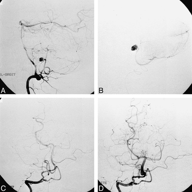
Case 1: 39-year-old man. A, Right vertebral injection, lateral view, shows aneurysm located at the telovelotonsillar segment (cranial loop) bifurcation of the PICA in its hemispheric and vermian branches. B, Intraaneurysmal contrast injection, lateral view, shows opacification of the aneurysm and progressive filling of the hemispheric branch. C, Postembolization right vertebral injection, anteroposterior view, shows occlusion of the aneurysm with a small remnant at the level of the neck. D, 7-month follow-up right vertebral injection, anteroposterior view, shows complete occlusion of the aneurysm, with no visible neck remnant. The distal PICA appears normal.
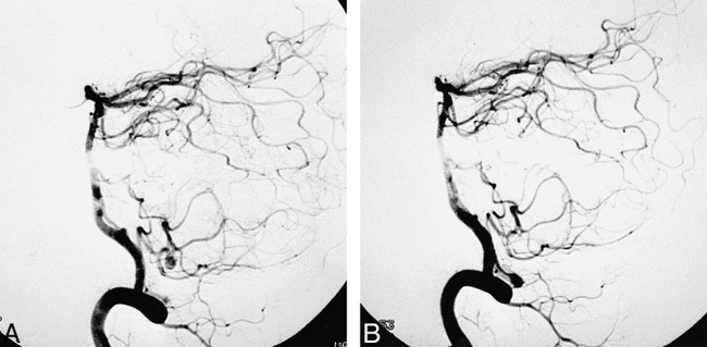
Case 2: 38-year-old woman. A, Right vertebral injection, lateral view, shows aneurysm located at the telovelotonsillar segment bifurcation of the PICA, in its hemispheric and vermian branches. B, Postembolization right vertebral injection, lateral view, shows complete occlusion of the aneurysm with normal-appearing distal PICA.
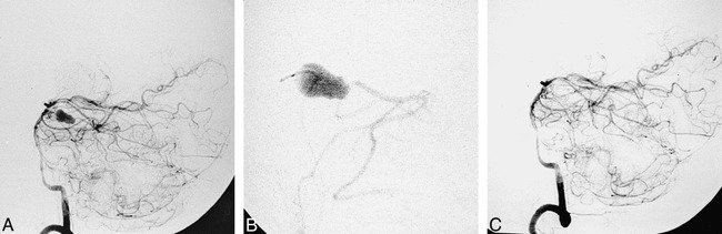
Case 3: 44-year-old woman. A, Left vertebral injection, lateral view, shows distal aneurysm in the hemispheric branch of the right SCA. B, Intraaneurysmal contrast injection, lateral view, shows opacification of the aneurysm. Note absence of an aneurysmal neck and filling of the distal branch. C, Postembolization left vertebral injection, lateral view, shows occlusion of the aneurysm and parent artery. Proximal SCA segment is not opacified owing to spasm caused by withdrawal of the microcatheter.
References
-
- Locksley HB. Report on the cooperative study of intracranial aneurysms and subarachnoid hemorrhage, section V, part 1: natural history of subarachnoid hemorrhage, intracranial aneurysms and arterio-venous malformations; based on 6368 cases in the cooperative study. J Neurosurg 1966;25:219-239 - PubMed
-
- Gacs G, Vinuela F, Fox AJ, Drake CG. Peripheral aneurysms of the cerebellar arteries: review of 16 cases. J Neurosurg 1983;58:63-68 - PubMed
-
- Nishizaki T, Tamaki N, Nishida Y, Fujita K, Matsumoto S. Aneurysms of the distal postero inferior cerebellar artery: experience with three cases and review of the literature. Neurosurgery 1985;16:829-832 - PubMed
-
- Spallone A, De Santis S, Giuffre R. Peripheral aneurysms of the anterior inferior cerebellar artery: case report and review of literature. Br J Neurosurg 1995;9:537-541 - PubMed
-
- Andoh T, Itoh T, Yoshimura S,, et al. Peripheral aneurysms of the posterior inferior cerebellar artery: analysis of 15 cases. No Shinkei Geka 1992;20:683-690 - PubMed
Publication types
MeSH terms
Substances
LinkOut - more resources
Full Text Sources
Other Literature Sources
Medical