

Human right and left colon differ in epithelial cell apoptosis and in expression of Bak, a pro-apoptotic Bcl-2 homologue
- PMID: 10369703
- PMCID: PMC1727558
- DOI: 10.1136/gut.45.1.45
Human right and left colon differ in epithelial cell apoptosis and in expression of Bak, a pro-apoptotic Bcl-2 homologue
Abstract
Background: Propensity to colonic neoplasia differs between the right and left colon.
Aims: To examine whether this difference may be related to regional differences in epithelial apoptosis, in expression of a proapoptotic regulatory protein, Bak, and in proliferation.
Patients: Individuals with no history of colorectal neoplasia.
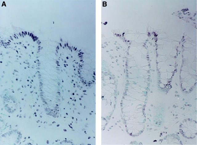
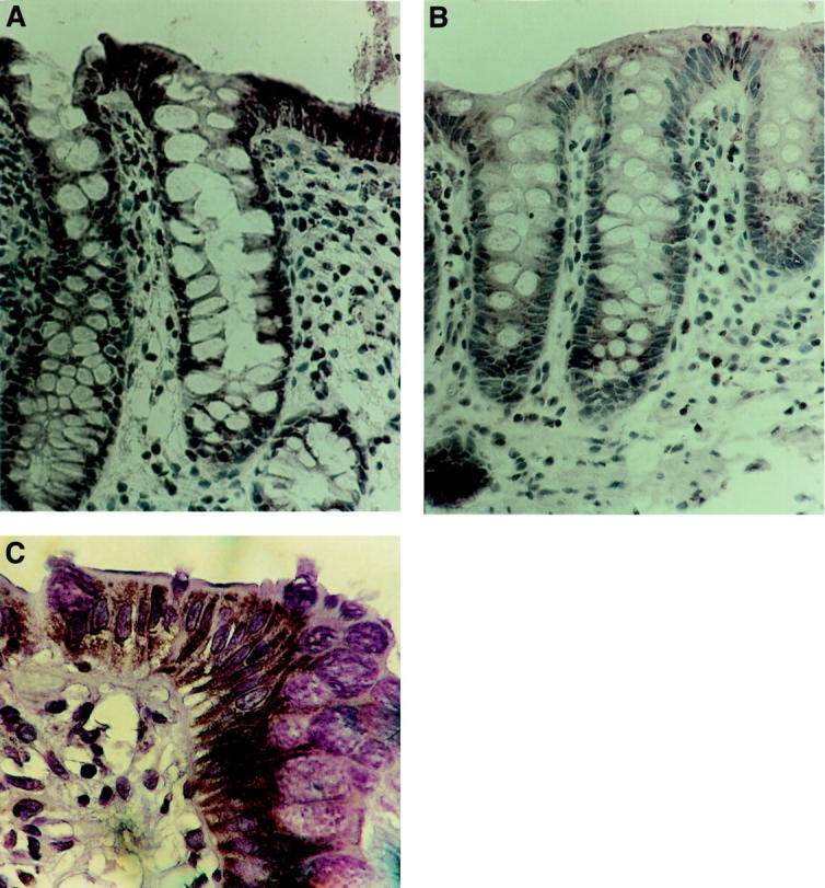
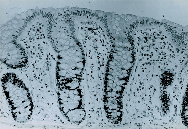
Methods: Archival blocks of colorectal tissues were immunostained for proliferating cells (antibody to Ki-67 antigen), and Bak expression (polyclonal antiserum). Cells containing DNA strand breaks, a marker of apoptosis, were identified by terminal deoxyuridine nucleotidyl nick end labelling (TUNEL).
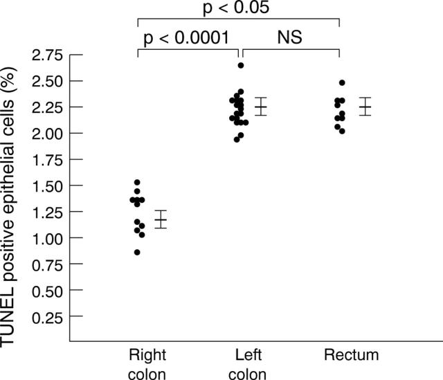
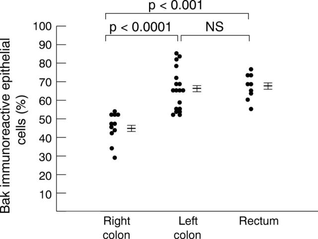
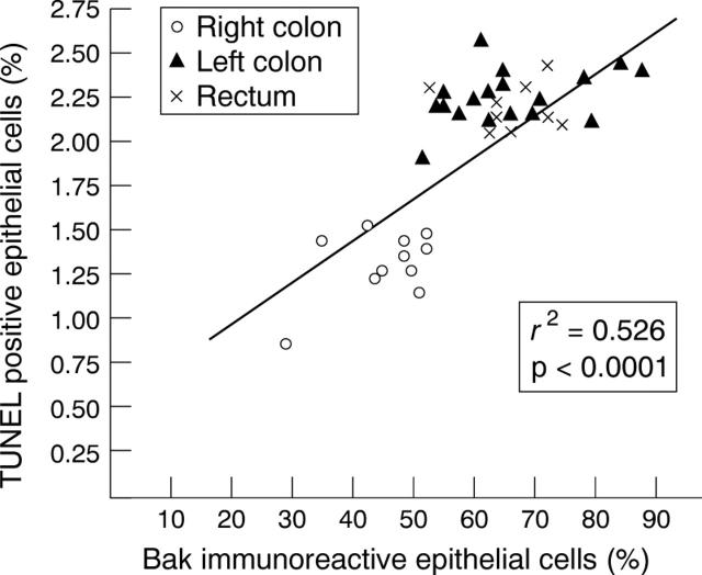
Results: There were fewer TUNEL positive epithelial cells in the right colon (mean 1.2 (SE 0.1)% of all epithelial cells) than the left colon (2.2 (0.1)%, p<0.0001) or rectum (2.2 (0.3)%, p<0.05). Bak expression was less common in the right colon (mean 46 (2.3)% of epithelial cells immunoreactive) than the left colon (66 (2.7)%, p<0.0001), or rectum (67 (2.3)%, p<0.001). Bak expression and TUNEL positivity were highly positively correlated (p<0.0001). In contrast to apoptosis, mean whole crypt proliferation labelling index was similar throughout the colorectum (right colon: 15.6 (3.2)%; left colon: 13. 5 (1.2)%; rectum: 13.3 (2.3)%).
Conclusion: The percentage of proliferating colonic epithelial cells is constant throughout the colon, but fewer epithelial cells undergo Bak mediated apoptosis in the right than in the left colon or rectum. This suggests that colonocytes may be lost by methods other than apoptosis in the right colon.
Figures
References
MeSH terms
Substances
LinkOut - more resources
Full Text Sources