. 1999 Apr;126(8):1847-55.
doi: 10.1038/sj.bjp.0702479.

Direct inhibition of the N-methyl-D-aspartate receptor channel by dopamine and (+)-SKF38393

Affiliations

Direct inhibition of the N-methyl-D-aspartate receptor channel by dopamine and (+)-SKF38393

N G Castro et al. Br J Pharmacol. 1999 Apr.

Abstract

1. Dopamine is known to modulate glutamatergic synaptic transmission in the retina and in several brain regions by activating specific G-protein-coupled receptors. We have examined the possibility of a different type of mechanism for this modulation, one involving direct interaction of dopamine with ionotropic glutamate receptors. 2. Ionic currents induced by fast application of N-methyl-D-aspartate (NMDA) were recorded under whole-cell patch-clamp in cultured striatal, thalamic and hippocampal neurons of the rat and in retinal neurons of the chick. Dopamine at concentrations above 100 microM inhibited the NMDA response in all four neuron types, exhibiting an IC50 of 1.2 mM in hippocampal neurons. The time course of this inhibition was fast, developing in less than 100 ms. 3. The D1 receptor agonist (+)-SKF38393 mimicked the effect of dopamine, with an IC50 of 58.9 microM on the NMDA response, while the enantiomer (-)-SKF38393 was ineffective at 50 microM. However, the D1 antagonist R(+)-SCH23390 did not prevent the inhibitory effect of (+)-SKF38393. 4. The degree of inhibition by dopamine and (+)-SKF38393 depended on transmembrane voltage, increasing 2.7 times with a hyperpolarization of about 80 mV. The voltage-dependent block by dopamine was also observed in the presence of MgCl2 1 mM. 5. Single-channel recordings showed that the open times of NMDA-gated channels were shortened by (+)-SKF38393. 6. These data suggested that the site to which the drugs bound to produce the inhibitory effect was distinct from the classical D1-type dopamine receptor sites, possibly being located inside the NMDA channel pore. It is concluded that dopamine and (+)-SKF38393 are NMDA channel ligands.


Figures

Figure 1
Figure 1
Inhibition by dopamine of NMDA-gated currents. (a) NMDA 10 μM plus glycine 10 μM applied in a 3-s pulse (open bar) to a striatal neuron clamped at −57 mV elicited an inward current that quickly decayed to about 25% of the peak amplitude. When the pulse was applied in the presence of dopamine 100 or 500 μM, the peak current amplitude was reduced. (b and c) A 6-s pulse of NMDA 10 μM applied to a rat hippocampal (b) or a chick retinal neuron (c) clamped at −57 mV elicited slowly-decaying inward currents that were reversibly reduced when dopamine 1 mM was co-applied for 2 s, in the middle of the NMDA pulse. The horizontal dashed line indicates the baseline current level and the long and short open bars on top of the traces indicate the durations of the agonist (NMDA) and antagonist (dopamine) pulses, respectively. The vertical calibration bars correspond to 50 pA in (a, b and c).
Figure 2
Figure 2
(a) Inhibition of currents elicited by NMDA 10 μM and glycine 10 μM by (+)-SKF38393. Five traces obtained from the same hippocampal neuron were scaled to the same peak amplitude and superimposed, illustrating the control and the effect of the indicated concentrations of (+)-SKF38393 in micromolar units. The peak amplitude of the NMDA current ranged from −100 to −150 pA in this cell. (b) Inhibition curve of the NMDA-gated currents by dopamine and (+)-SKF38393 in hippocampal neurons clamped at −57 mV. Each symbol and error bar mark the mean±s.e.mean of 3–8 neurons. The best-fitting curves were obtained with IC50 and nH values of 1.2 mM and 1.16 for dopamine, and of 58.9 μM and 1.21 for (+)-SKF38393.
Figure 3
Figure 3
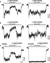
Stereoselectivity and specificity of the inhibition of the NMDA currents by (+)-SKF38393. Currents activated by NMDA 50 μM and glycine 10 μM were blocked by the (+), but not by the (−) isomer of SKF38393 (50 μM) when tested in the same neuron of the chick retina (a) or rat thalamus (b). In another thalamic neuron (c), (+)-SKF38393 50 μM reduced the NMDA-gated current to 48.5%, but the non-desensitizing current activated by kainate (50 μM) was unaffected.
Figure 4
Figure 4
Voltage-dependence of the blocking effect in hippocampal neurons. (a) Currents activated by NMDA 10 μM and glycine 10 μM with a superimposed pulse of dopamine 1 mM in one hippocampal neuron held at +33, +3, −27, −57 and −87 mV were scaled and superimposed to show that the degree of current blockade increases with hyperpolarization. (b) Plots of NMDA current in dopamine vs membrane potential. The data points obtained in the absence of Mg2+ are from the experiment in (a), and are shown with the best-fitted exponential curve. The mean data points from two experiments done in the presence of MgCl2 1 mM also lie close to the curve. (c) Traces from another hippocampal neuron to which NMDA and (+)-SKF38393 50 μM were applied at −37, −57, −77, and −97 mV, scaled as in (a). (d) Plot of current in (+)-SKF38393 vs membrane potential (means±s.d.) from four cells, including that in (c). The data from individual cells and the best-fitting exponential are also shown.
Figure 5
Figure 5
Effect of (+)-SKF38393 on channel open time in hippocampal neurons. Single-channel currents activated by NMDA 50 μM and glycine 10 μM in one outside-out membrane patch (a) appeared shorter when (+)-SKF38393 50 μM was added (b). Open-time histograms from these recordings could be fitted with simple exponential distribution functions. With NMDA and glycine alone (c), the mean open time was 2.43 ms, while in (+)-SKF38393 (d), it was 0.76 ms. The patch was held at −87 mV. Closed and open-channel current levels are labeled c and o, respectively, in panel (a).

Similar articles

Cited by

References

    1. AKAIKE A., OHNO Y., SASA M., TAKAORI S. Excitatory and inhibitory effects of dopamine on neuronal activity of the caudate nucleus neurons in vitro. Brain Res. 1987;418:262–272. - PubMed
    1. ALKONDON M., ALBUQUERQUE E.X. Diversity of nicotinic acetylcholine receptors in rat hippocampal neurons. I. Pharmacological and functional evidence for distinct structural subtypes. J. Pharmacol. Exper. Therap. 1993;265:1455–1473. - PubMed
    1. ANDERSEN P.H., JANSEN J.A. Dopamine receptor agonists: selectivity and dopamine D1 receptor efficacy. Eur. J. Pharmacol. 1990;188:335–347. - PubMed
    1. ASCHER P., NOWAK L. The role of divalent cations in the N-methyl-d-aspartate responses of mouse central neurones in culture. J. Physiol. (London) 1988;399:247–266. - PMC - PubMed
    1. BRAUN A., FABBRINI G., MOURADIAN M.M., SERRATI C., BARONE P., CHASE T.N. Selective D-1 dopamine receptor agonist treatment of Parkinson's disease. J. Neural Transm. 1987;68:41–50. - PubMed

Publication types

MeSH terms

LinkOut - more resources