. 1999 Jul;19(7):4582-91.
doi: 10.1128/MCB.19.7.4582.

Dominant negative murine serum response factor: alternative splicing within the activation domain inhibits transactivation of serum response factor binding targets

Affiliations

Dominant negative murine serum response factor: alternative splicing within the activation domain inhibits transactivation of serum response factor binding targets

N S Belaguli et al. Mol Cell Biol. 1999 Jul.

Abstract

Primary transcripts encoding the MADS box superfamily of proteins, such as MEF2 in animals and ZEMa in plants, are alternatively spliced, producing several isoformic species. We show here that murine serum response factor (SRF) primary RNA transcripts are alternatively spliced at the fifth exon, deleting approximately one-third of the C-terminal activation domain. Among the different muscle types examined, visceral smooth muscles have a very low ratio of SRFDelta5 to SRF. Increased levels of SRFDelta5 correlates well with reduced smooth muscle contractile gene activity within the elastic aortic arch, suggesting important biological roles for differential expression of SRFDelta5 variant relative to wild-type SRF. SRFDelta5 forms DNA binding-competent homodimers and heterodimers. SRFDelta5 acts as a naturally occurring dominant negative regulatory mutant that blocks SRF-dependent skeletal alpha-actin, cardiac alpha-actin, smooth alpha-actin, SM22alpha, and SRF promoter-luciferase reporter activities. Expression of SRFDelta5 interferes with differentiation of myogenic C2C12 cells and the appearance of skeletal alpha-actin and myogenin mRNAs. SRFDelta5 repressed the serum-induced activity of the c-fos serum response element. SRFDelta5 fused to the yeast Gal4 DNA binding domain displayed low transcriptional activity, which was complemented by overexpression of the coactivator ATF6. These results indicate that the absence of exon 5 might be bypassed through recruitment of transcription factors that interact with extra-exon 5 regions in the transcriptional activating domain. The novel alternatively spliced isoform of SRF, SRFDelta5, may play an important regulatory role in modulating SRF-dependent gene expression.


Figures
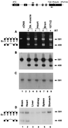
FIG. 1
FIG. 1
Alternative splicing of SRF RNA removes exon 5. The exon-intron organization of the SRF genome and positions of the primers used for PCR are shown at the top. (A) Ethidium bromide-stained agarose gel showing the RT-PCR products from skeletal muscle (lanes 3 and 4), heart (lanes 5 and 6), brain (lane 7 and 8), and 10T1/2 cells (lane 9). Cloned mSRF cDNA was used as the template in lane 1. Lane 2 contained no input cDNA. RT was omitted during cDNA synthesis for lanes 4, 6, and 8. Constitutively spliced and alternatively spliced forms are diagrammatically represented on the left, and sizes of the bands are shown in base pairs on the right. Southern blots of the gel probed with the mSRF cDNA and exon 5 oligonucleotide are shown in panels B and C, respectively. End-labeled primers were used for PCR shown in panel D.
FIG. 2
FIG. 2
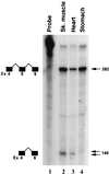
Tissue-restricted expression of alternatively spliced SRFΔ5 RNA, determined by RNase protection analysis of total cellular RNA isolated from skeletal muscle (lane 2), heart (lane 3), and stomach (lane 4) with the antisense SRF probe. The undigested probe was run in lane 1. Protected unspliced and spliced products are diagrammatically represented to the left; sizes of the protected fragments are indicated in base pairs on the right.
FIG. 3
FIG. 3
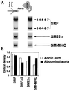
Inverse gradient of expression of SRFΔ5, SM22α, and SM-MHC along the aorta. (A) Total RNA isolated from the aortic arch (lane 1) and abdominal aorta (lane 2) analyzed by semiquantitative RT-PCR with the indicated end-labeled primers. SRF was amplified for 25 cycles, and SM22α and SM-MHC were amplified for 20 cycles. The linearity of amplification was confirmed by harvesting the amplified products at 20, 23, 25, 28, and 30 cycles. Constitutively and alternatively spliced isoforms of SRF are diagrammatically represented to the right. (B) Relative optical densities of bands.
FIG. 4
FIG. 4
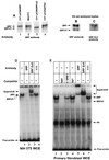
SRFΔ5 protein is expressed in NIH 3T3 cells and several other mouse cell lines. (A to C) Western blot analysis of SRF and SRFΔ5 proteins. Protein extracts (20 μg) prepared from NIH 3T3 cells and day 4 EBs were used for Western analysis. NIH 3T3 cell extract (lane 2) was mixed with CV1 whole-cell extract overexpressing SRF (lane 1), SRFΔ5 (lane 3), and GATA-4 (lane 4) and probed with the polyclonal immune serum raised against bacterially expressed SRF protein. Lanes 2 and 4 were exposed three to four times longer than lanes 1 and 3. In panel B, the C-terminal epitope-specific antibody (Santa Cruz) was used. This blot was stripped and subsequently reacted with an exon 5 epitope-specific immune serum (C). Expression of SRFΔ5 proteins in NIH 3T3 cells (D) and mouse embryonic fibroblasts (E) was demonstrated by EMSA. Whole-cell extracts (WCE; 5 μg) were preincubated with a 50-fold excess of the indicated specific (self and cardiac α-actin SRE1) and nonspecific (Sp1) competitors and 0.5 μl of polyclonal immune serum. Annealed SRE1 oligonucleotide from the SRF promoter was the probe. Positions of SRF, SRFΔ5, and supershifted and nonspecific (NS) complexes and of the free probe are indicated. The gel in panel E was run for a longer time to resolve the SRF and SRFΔ5 complexes.
FIG. 5
FIG. 5
SRFΔ5 forms DNA binding-competent homodimers and heterodimers. (A) The EMSA conditions were as described for Fig. 4D and E. The binding reaction mixture contained unprogrammed (UP) RRL (lane 2), 2.5 μl of in vitro-translated SRFΔ5 in programmed (P) RRL (lanes 3 and 5), and 2.5 μg of CV1 whole-cell extract overexpressing SRFΔC (lanes 4 and 5). In vitro-translated SRFΔ5 was preincubated with CV1 whole-cell extract overexpressing SRFΔC in lane 5. SRFΔ5 and SRFΔC homodimers and SRFΔ5-SRFΔC heterodimers are indicated by arrows to the left. (B) SRFΔ5 heterodimerizes with SRF independent of DNA binding. [35S]methionine-labeled in vitro-translated luciferase and SRFΔ5 were incubated with GST (lanes 2 and 5) or GST-SRF (lanes 3 and 6) immobilized on glutathione beads, washed extensively, and analyzed on an SDS–10% polyacrylamide gel. Lane 1 and 4 contained 10% of the input luciferase and SRFΔ5, respectively.
FIG. 6
FIG. 6
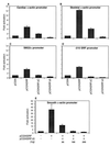
SRFΔ5 inhibits SRE-dependent promoter activity. Subconfluent CV1 cells were cotransfected with 1 μg of cardiac α-actin (A), skeletal α-actin (B), SM22α (C) and −310 SRF (D) promoter-luciferase reporter plasmids and 150 ng of expression vectors for SRF (pCGNSRF), SRFΔ5 (pCGNSRFΔ5), C-terminally truncated mutant of SRF (pCGNSRFΔC), or the empty vector pCGN. For panel E, 200 ng of smooth α-actin reporter was cotransfected with 150 ng of pCGNSRF or a combination of pCGNSRF and the indicated amounts of pCGNSRFΔ5. Cells were harvested 48 h posttranscription, and the luciferase activity was measured. Results shown are mean ± standard error of the mean for three duplicate experiments.
FIG. 7
FIG. 7
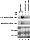
SRFΔ5 inhibits differentiation of myogenic C2C12 cells. C2C12 cells were cotransfected with SRFΔ5 and SRFpm1 expression plasmids and pSV2neo vector. G418-resistant cell populations were grown to 50% confluence in growth medium (GM) and induced to differentiate for 3 days by adding differentiation medium (DM). Total RNA (20 μg) was subjected to Northern analysis to detect expression of the skeletal muscle-specific factors skeletal α-actin (αSk actin) and myogenin, using random-primed probes. 28S and 18S rRNA bands were visualized by ethidium bromide staining to show RNA loading. Expression of skeletal α-actin and myogenin was significantly inhibited in both mutant cell lines.
FIG. 8
FIG. 8
SRFΔ5 represses serum-induced activity of c-fos SRE. 10T1/2 murine fibroblasts were transfected with 200 ng each of the internal control plasmid pCMVβGal, luciferase reporter plasmid c-fos SRELuc, and expression plasmids pCGNSRF, pCGNSRFΔ5, pCGNSRFΔC or the empty vector pCGN. After transfection, cells were maintained in DMEM containing 10% serum for 16 h. Cells were serum starved for 48 h in DMEM containing 0.5% serum and then induced with 20% serum for 3 h. Luciferase activity was normalized to β-galactosidase activity. Similar results were obtained for NIH 3T3 cells.
FIG. 9
FIG. 9
ATF6 complements Gal4-SRF C-terminal Δ5 transcriptional activity. CV1 cells were transfected with 1 μg of the Gal4 luciferase reporter (G5luc), Gal4DB, Gal4SRF 266-508, SRFΔ5, and ATF6 expression vectors in a variety of combinations as indicated. Cells were harvested 48 h posttransfection, and luciferase activity was measured. The results shown are from three experiments.

Similar articles

Cited by

References

    1. Annweiler A, Muller-Immergluck M, Wirth T. Oct2 transactivation from a remote enhancer position requires a B-cell-restricted activity. Mol Cell Biol. 1992;12:3107–3116. - PMC - PubMed
    1. Arsenian S, Weinhold B, Oelgeschlager M, Ruther U, Nordheim A. Serum response factor is essential for mesoderm formation during mouse embryogenesis. EMBO J. 1998;17:6289–6299. - PMC - PubMed
    1. Belaguli N S, Schildmeyer L A, Schwartz R J. Organization and myogenic restricted expression of the murine serum response factor gene. J Biol Chem. 1997;272:18222–18231. - PubMed
    1. Belaguli, N. S., J. L. Sepulveda, V. Nigam, and R. J. Schwartz. Unpublished data.
    1. Bickmore W A, Oghene K, Little M H, Seawright A, VanHeyningen V, Hastie N D. Modulation of DNA binding specificity by alternative splicing of the Wilms tumor wt1 gene transcript. Science. 1992;257:235–238. - PubMed

Publication types

MeSH terms

LinkOut - more resources