. 1999 Jul;19(7):4611-22.
doi: 10.1128/MCB.19.7.4611.

Ras-specific exchange factor GRF: oligomerization through its Dbl homology domain and calcium-dependent activation of Raf

Affiliations

Ras-specific exchange factor GRF: oligomerization through its Dbl homology domain and calcium-dependent activation of Raf

P H Anborgh et al. Mol Cell Biol. 1999 Jul.

Abstract

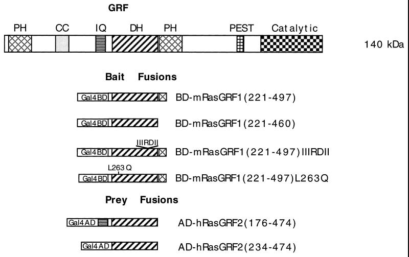
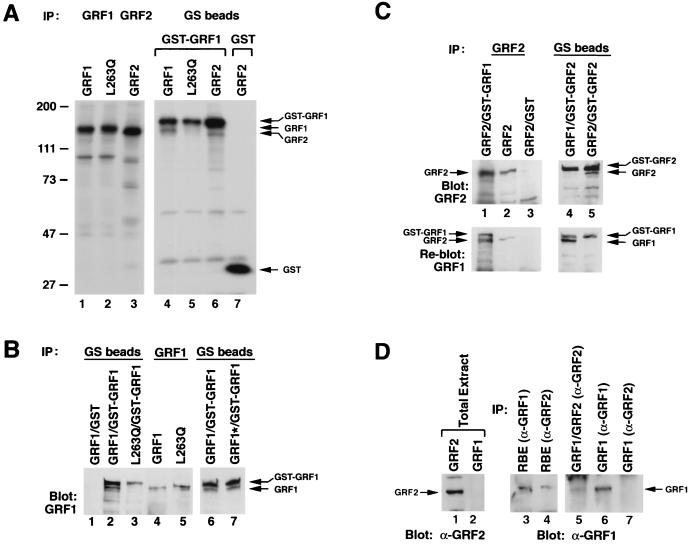
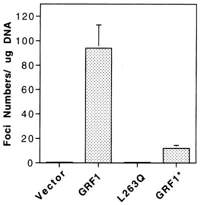
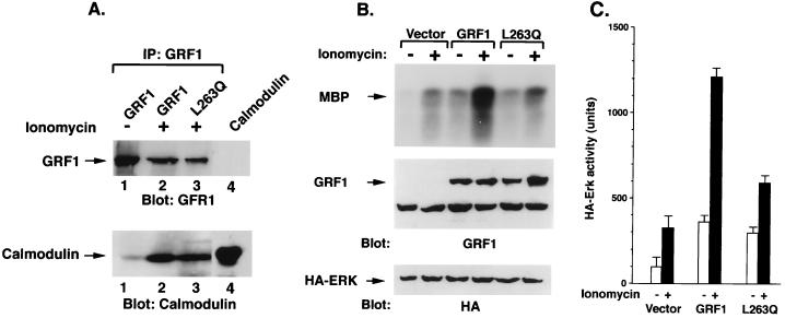
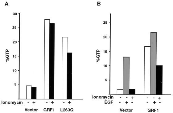
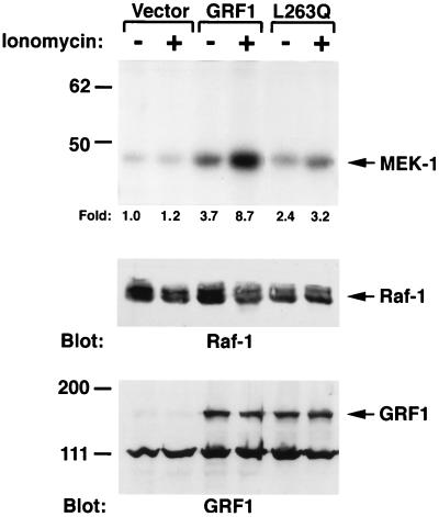
The full-length versions of the Ras-specific exchange factors Ras-GRF1 (GRF1) and Ras-GRF2 (GRF2), which are expressed in brain and a restricted number of other organs, possess an ionomycin-dependent activation of Erk mitogen-activated protein kinase activity in 293T cells (C. L. Farnsworth et al., Nature 376:524-527, 1995; N. P. Fam et al., Mol. Cell. Biol. 17:1396-1406, 1996). Each GRF protein contains a Dbl homology (DH) domain. A yeast two-hybrid screen was used to identify polypeptides that associate with the DH domain of GRF1. In this screen, a positive cDNA clone from a human brain cDNA library was isolated which consisted of the GRF2 DH domain and its adjacent ilimaquinone domain. Deletion analysis verified that the two-hybrid interaction required only the DH domains, and mutation of Leu-263 to Gln (L263Q) in the N terminus of the GRF1 DH domain abolished the two-hybrid interaction, while a cluster of more C-terminally located mutations in the DH domain did not eliminate the interaction. Oligomers between GRF1 and GRF2 were detected in a rat brain extract, and forced expression of GRF1 and GRF2 in cultured mammalian cells formed homo- and hetero-oligomers. Introduction of the L263Q mutation in GRF1 led to a protein that was deficient in oligomer formation, while GRF1 containing the DH cluster mutations formed homo-oligomers with an efficiency similar to that of wild type. Compared to wild-type GRF1, the focus-forming activity on NIH 3T3 cells of the GRF1 DH cluster mutant was reduced, while the L263Q mutant was inactive. Both mutants were impaired in their ability to mediate ionomycin-dependent Erk activity in 293T cells. In the absence of ionomycin, 293T cells expressing wild-type GRF1 contained much higher levels of Ras-GTP than control cells; the increase in Erk activity induced by ionomycin in the GRF1-expressing cells also induced a concomitant increase in Raf kinase activity, but without a further increase in the level Ras-GTP. We conclude that GRF1 and GRF2 can form homo- and hetero-oligomers via their DH domains, that mutational inactivation of oligomer formation by GRF1 is associated with impaired biological and signaling activities, and that in 293T cells GRF1 mediates at least two pathways for Raf activation: one a constitutive signal that is mainly Ras-dependent, and one an ionomycin-induced signal that cooperates with the constitutive signal without further augmenting the level of GTP-Ras.


Figures

FIG. 1
FIG. 1
Schematic representation of the domain structure of GRF1 and of the baits and preys used in the two-hybrid interactions. The various abbreviations are as defined in the text; PEST is the protein instability motif, and Catalytic refers to the Ras guanine nucleotide exchange domain. In the bait fusions, Gal4BD represents the Gal4-binding domain (BD), while Gal4AD represents the Gal4 activation domain (AD) in the prey fusions. The numbers in parentheses refer to the corresponding amino acid residues of full-length mGRF1 encoded in the bait fusions and of full-length hGRF2 in the prey fusions. BD-mGRF1(221–497) is the starting bait fusion used in the two-hybrid screen. BD-mGRF1(221–460) lacks the PH domain codons in BD-mGRF1(221–497). BD-mGRF(221–497)IIIRDII harbors a cluster of substitutions in codons 394 to 400, while BD-mGRF1(221–497)L263Q contains a point mutation (see the text). AD-hGRF2(176–474) is the insert from clone pGAD10.56 obtained in the two-hybrid screen. In AD-hGRF2(234–474), the IQ motif codons have been deleted from the hGRF2 insert obtained in the two-hybrid screen.
FIG. 2
FIG. 2
Comparison of the sequences of mouse (m) and human (h) forms of Ras-GRF1 (GRF1) and Ras-GRF2 (GRF2). Alignment is given in comparison with hRas-GRF2 (hGRF2). Going from the N terminus to the C terminus, the boxes indicate the PH, IQ, DH-PH, and the Ras catalytic domains, respectively (see Fig. 1). An asterisk indicates sequence identity with respect to hRas-GRF2; a dash indicates a deletion. The underlined sequence of mRas-GRF1 (mGRF1) corresponds to the bait used in the two-hybrid screen (residues 221 to 497 of mGRF1). The underlined sequence of hGRF2 corresponds to the insert of clone pGAD10.56, the sequence trapped in the two-hybrid screen (residues 176 to 474 of hRas-GRF2). The GenBank accession numbers are as follows: L20899 (mGRF1), U67326 (mGRF2), L26584 (hGRF1), and AF023130 (hGRF2). The following regions are not highly conserved between GRF1 and GRF2: (i) the most N-terminal PH domain (and to a lesser extent the PH domain C terminal to the DH domain); (ii) the region corresponding to residues Ser-180–Glu-200 of hGRF2, in which only 6 of 21 residues are identical between mGRF1 and hGRF2 (this region was used to generate a GRF2-specific peptide antibody); and (iii) the region corresponding to residues Thr761–Pro-878 of hGRF2, in which only 20 of 118 residues are identical between hGRF2 and hGRF1. This region, which is much shorter in mGRF2, was chosen to generate cDNA probes for Northern analysis of human tissues.
FIG. 3
FIG. 3
Formation of GRF1 and GRF2 oligomers in mammalian cells. (A) GRF oligomerization occurs in NIH 3T3 cells between GST-GRF1 and wild-type GRF1 or wild-type GRF2, but it is deficient between GST-GRF1 and the L263Q GRF1 mutant. Metabolically labeled NIH 3T3 cells transiently expressed mGRF1, mGRF1L263Q, and hGRF2, with or without GST-mGRF1 or GST, as indicated. Cell lysates were immunoprecipitated with antisera to GRF1 (lanes 1 and 2), GRF2 (lane 3) or precipitated with glutathione-Sepharose (GS) beads (lanes 4 to 7). The arrows indicate the location of GRF1 (lanes 4 and 5) or GRF2 (lane 6) coprecipitated with GST-GRF1 (lanes 4 to 6) and the location of GST (lane 7). (B) Wild-type GRF1 and the DH cluster mutant form oligomers in 293T cells, but oligomerization is deficient with the L263Q mutant. Transiently transfected cells expressing wild-type (lanes 1, 2, 4, and 6) or mutant GRF1 (L263Q in lanes 3 and 5; cluster mutant [GRF1*] in lane 7), with or without coexpression of GST-GRF1, were precipitated by glutathione-Sepharose (GS) beads or GRF antibodies as indicated, followed by anti-GRF1 blotting. The designated oligomerized proteins are marked with arrows. (C) Oligomerization in 293T cells. In lanes 1 to 3, lysates from cells expressing GRF2 with or without coexpression of GST-GRF1 or GST were immunoprecipitated and blotted with anti-GRF2. In lanes 4 and 5, lysates from cells coexpressing GST-GRF2 and GRF1 or GRF2 were precipitated with glutathione-Sepharose (GS) beads followed by anti-GRF2 blotting. In the bottom panels, the anti-GRF2 blots were (incompletely) stripped and were reprobed with GRF1 antibodies. The designated oligomerized proteins are marked with arrows. (D) A rat brain extract (RBE) contains GRF1-GRF2 oligomers. Lanes: 1 and 2, extracts from 293T cells expressing GRF2 and GRF1, respectively, Western blotted with anti-GRF2 antibodies; 3 and 4, rat brain extracts immunoprecipitated with anti-GRF1 and anti-GRF2, respectively; 5, 293T cells coexpressing GRF1 and GRF2 immunoprecipitated with anti-GRF2 antibodies; 6 and 7, 293T cells expressing GRF1 immunoprecipitated with anti-GRF1 or anti-GRF2 antibodies, respectively.
FIG. 4
FIG. 4
Focal transforming activity of GRF1. NIH 3T3 cells transfected with the indicated GRF1 constructs. The data shown represent the mean of four separate experiments (three readings per experiment). Each bar shows the transforming activity and the standard error.
FIG. 5
FIG. 5
Erk and GTP-Ras in NIH 3T3 cells. Assays were carried out 2 days after transfection of NIH 3T3 cells with the indicated GRF1 plasmid. (A) Erk activity in NIH 3T3 cells. Cells were transiently cotransfected with empty vector, wild-type GRF1 (GRF1), or the L263Q GFR1 mutant (L263Q), along with HA-ERK2. In the upper portion of the panel, the basal Erk activity was determined by precipitating lysates with HA antibody, followed by kinase assay of the immune complex with MBP as the substrate. In the middle portion of the panel, the presence of GRF1 or the mutant was verified by Western blotting of the immunoprecipitates. In the lower portion of the panel, the blots were stripped and probed with the HA antibody to verify the equal expression of HA-Erk2. The arrows show the location of MBP, GRF1, and HA-Erk in the upper, middle, and lower portions, respectively. (B) In vivo measurement of GTP and GDP bound to Ras in transiently transfected NIH 3T3 cells. Cells were metabolically labeled with [32P]orthophosphate, extracts were immunoprecipitated with a Ras-specific monoclonal antibody, and the percentage of GTP-Ras was determined by thin-layer chromatography as described in Materials and Methods.
FIG. 6
FIG. 6
Calmodulin binding and ERK activity in 293T cells expressing wild-type GRF1 and L263Q mutant. (A) 293T cells were transiently transfected with wild-type GRF1 or the L263Q mutant, serum starved overnight, and either left untreated (−) or treated with ionomycin for 5 min (+). Cell lysates were immunoprecipitated with GRF1 antibodies and blotted with GRF1 antibodies (top portion of the panel) or with a calmodulin antibody (bottom portion of panel). Lane 4 contains only input calmodulin marker. (B) Basal and ionomycin-induced Erk activation in 293T cells transfected with GRF1. Cells were transiently transfected with vector, wild-type GRF1, or the L263Q mutant along with HA-ERK2, serum starved overnight, and either left untreated (−) or treated with ionomycin for 5 min (+). The exogenous ERK activity was determined as described in the text. The cells were processed and analyzed as described for panel A. (C) Quantitation of ERK activity. The amount of radioactivity present in phosphorylated MBP was quantitated with a phosphorimager. The activity of exogenous Erk was based on the mean of two experiments.
FIG. 7
FIG. 7
GTP-Ras in 293T cells expressing wild-type GRF1 and L263Q mutant. Cells were transiently transfected with the indicated GRF1 gene. Cultures were serum starved and metabolically labeled with [32P]orthophopshate, cells were treated with 5 μM ionomycin or 20 nM EGF for 5 min, extracts were immunoprecipitated with a Ras-specific monoclonal antibody, and the percentage of GTP-Ras was determined by thin-layer chromatography as described in Materials and Methods. Results in panel A represent the average of two experiments, while the results in panel B are from a single experiment.
FIG. 8
FIG. 8
GRF1 increased Raf activity in response to ionomycin. Transiently transfected 293T cells were treated with or without ionomycin after serum starvation as in Fig. 6. Cell lysates were immunoprecipitated with Raf antibodies, and the immunocomplexes were assayed for in vitro kinase activity by using MEK-1 as a substrate (upper part of panel). The amount of radioactivity present in phosphorylated MEK-1 was determined with a phosphoimager, and the fold increase, compared with the untreated vector control, is shown underneath the upper part of the panel. The expression of Raf and GRF1 was confirmed by immunoblotting, as shown in the middle and bottom panels, respectively.

Similar articles

Cited by

References

    1. Altschul S F, Madden T L, Schaffer A A, Zhang J, Zhang Z, Miller W, Lipman D J. Gapped blast and PSI-Blast: an new generation of protein database search programs. Nucleic Acid Res. 1997;25:3389–3402. - PMC - PubMed
    1. Baouz S, Jacquet E, Bernardi A, Parmeggiani A. The N-terminal moiety of CDC25Mm, a GDP/GTP exchange factor of Ras proteins, controls the activity of the catalytic domain: modulation by calmodulin and calpain. J Biol Chem. 1997;272:6671–6676. - PubMed
    1. Boguski M S, McCormick F. Proteins regulating Ras and its relatives. Nature. 1993;366:643–654. - PubMed
    1. Bowtell D, Fu P, Simon M, Senior P. Identification of murine homologues of the Drosophila son of sevenless gene—potential activators of ras. Proc Natl Acad Sci USA. 1992;89:6511–6515. - PMC - PubMed
    1. Brambilla R, Gnesutta N, Minichiello L, White G, Roylance A J, Herron C E, Ramsey M, Wolfer D P, Cestari V, Rossi-Arnaud C, Grant S G N, Chapman P F, Lipp H P, Sturani E, Klein R. A role for the Ras signalling pathway in synaptic transmission and long-term memory. Nature. 1997;390:281–286. - PubMed

MeSH terms

Associated data