. 1999 Jul;19(7):4684-94.
doi: 10.1128/MCB.19.7.4684.

Osmotic shock inhibits insulin signaling by maintaining Akt/protein kinase B in an inactive dephosphorylated state

Affiliations

Osmotic shock inhibits insulin signaling by maintaining Akt/protein kinase B in an inactive dephosphorylated state

D Chen et al. Mol Cell Biol. 1999 Jul.

Abstract

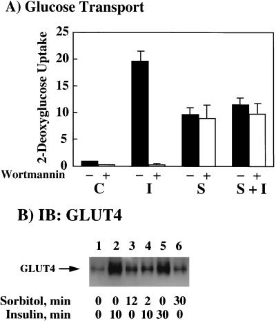
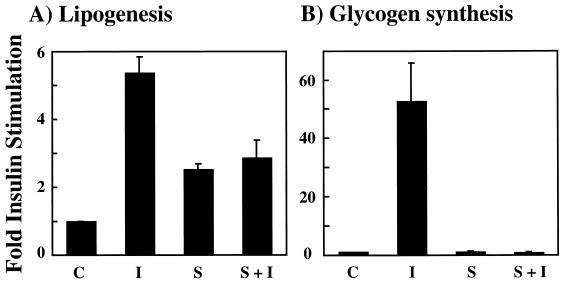
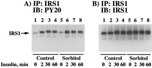
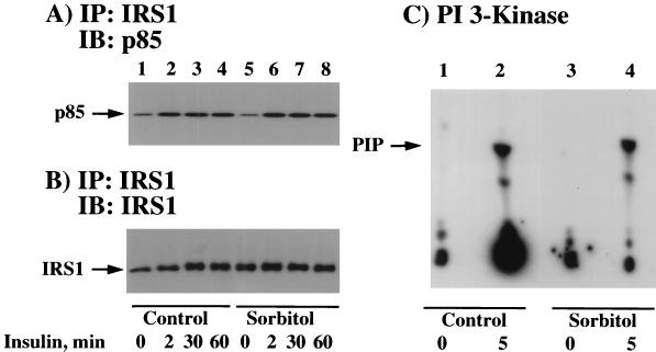
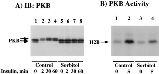
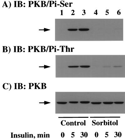
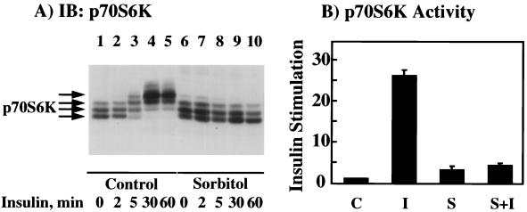
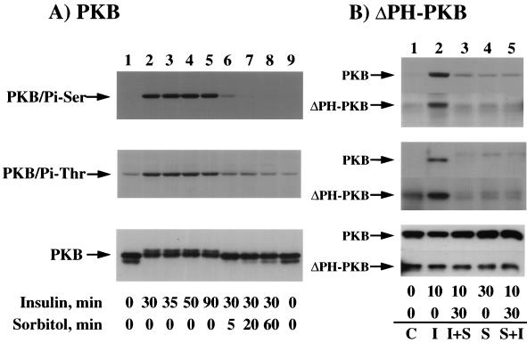
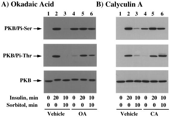
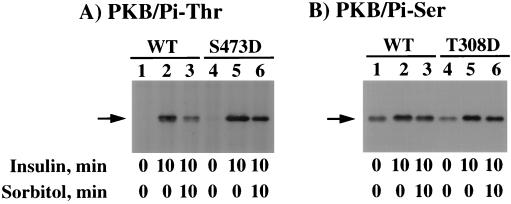
We have previously reported that insulin and osmotic shock stimulate an increase in glucose transport activity and translocation of the insulin-responsive glucose transporter isoform GLUT4 to the plasma membrane through distinct pathways in 3T3L1 adipocytes (D. Chen, J. S. Elmendorf, A. L. Olson, X. Li, H. S. Earp, and J. E. Pessin, J. Biol. Chem. 272:27401-27410, 1997). In investigations of the relationships between these two signaling pathways, we have now observed that these two stimuli are not additive, and, in fact, osmotic shock pretreatment was found to completely prevent any further insulin stimulation of glucose transport activity and GLUT4 protein translocation. In addition, osmotic shock inhibited the insulin stimulation of lipogenesis and glycogen synthesis. This inhibition of insulin-stimulated downstream signaling occurred without any significant effect on insulin receptor autophosphorylation or tyrosine phosphorylation of insulin receptor substrate 1 (IRS1). Furthermore, there was no effect on either the insulin-stimulated association of the p85 type I phosphatidylinositol (PI) 3-kinase regulatory subunit with IRS1 or phosphotyrosine antibody-immunoprecipitated PI 3-kinase activity. In contrast, osmotic shock pretreatment markedly inhibited the insulin stimulation of protein kinase B (PKB) and p70S6 kinase activities. In addition, the dephosphorylation of PKB was prevented by pretreatment with the phosphatase inhibitors okadaic acid and calyculin A. These data support a model in which osmotic shock-induced insulin resistance of downstream biological responses results from an inhibition of insulin-stimulated PKB activation.


Figures

FIG. 1
FIG. 1
Osmotic shock pretreatment inhibits insulin-stimulated glucose transport activity and GLUT4 translocation. (A) Differentiated 3T3L1 adipocytes were either left untreated (−, solid bars) or treated with 100 nM wortmannin (+, open bars) for 15 min at 37°C. The cells were then either left untreated (C), stimulated with 100 nM insulin for 15 min (I), incubated with 600 mM sorbitol for 45 min (S), or preincubated with 600 mM sorbitol for 30 min and then stimulated with insulin for another 15 min (S+I). The initial rate of 2-[3H]deoxyglucose uptake was then determined as described in Materials and Methods. The basal rate of 2-[3H]deoxyglucose uptake activity was normalized to 1. Error bars indicate standard deviations. (B) Differentiated 3T3L1 adipocytes were either left untreated (lane 1) or stimulated with 100 nM insulin for 10 (lane 2) or 30 (lane 5) min at 37°C. The 3T3L1 adipocytes were also pretreated with 600 mM sorbitol for 12 (lane 3) and 30 (lane 6) min at 37°C. In parallel, the cells were also pretreated with 600 mM sorbitol for 2 min and then incubated for an additional 10 min with 100 nM insulin (lane 4). Purified plasma membrane sheets were then prepared as described in Materials and Methods. The isolated plasma membranes were then immunoblotted (IB) with the GLUT4 antibody.
FIG. 2
FIG. 2
Osmotic shock pretreatment inhibits the insulin stimulation of lipogenesis and glycogen synthesis. Differentiated 3T3L1 adipocytes were either left untreated (C), stimulated with 100 nM insulin for 15 min (I), incubated with 600 mM sorbitol for 15 min (S), or preincubated with 600 mM sorbitol for 15 min and then stimulated with insulin for another 15 min (S+I). The initial rate of [14C]glucose incorporation into lipid (A) or glycogen (B) was determined as described in Materials and Methods. The basal rates of lipogenesis and glycogen synthesis were normalized to 1. Error bars indicate standard deviations.
FIG. 3
FIG. 3
Osmotic shock pretreatment does not affect insulin-induced tyrosine phosphorylation of the insulin receptor. Differentiated 3T3L1 adipocytes were either left untreated (lanes 1 to 4) or pretreated with 600 mM sorbitol (lanes 5 to 8) for 30 min at 37°C. The cells were then incubated in the absence of insulin (lanes 1 and 5) or in the presence of 100 nM insulin for 2 (lanes 2 and 6), 30 (lanes 3 and 7), or 60 (lanes 4 and 8) min. Whole-cell extracts were then prepared, and the insulin receptor (IR) was immunoprecipitated (IP) with an insulin receptor antibody as described in Materials and Methods. The immunoprecipitates were then immunoblotted (IB) with the PY20 phosphotyrosine antibody (A) or the insulin receptor antibody (B).
FIG. 4
FIG. 4
Osmotic shock pretreatment does not affect insulin-induced tyrosine phosphorylation of IRS1. Differentiated 3T3L1 adipocytes were either left untreated (lanes 1 to 4) or pretreated with 600 mM sorbitol (lanes 5 to 8) for 30 min at 37°C. The cells were then incubated in the absence of insulin (lanes 1 and 5) or in the presence of 100 nM insulin for 2 (lanes 2 and 6), 30 (lanes 3 and 7), or 60 (lanes 4 and 8) min. Whole-cell extracts were then prepared, and IRS1 was immunoprecipitated with an IRS1 antibody as described in Materials and Methods. The immunoprecipitates were then immunoblotted with the PY20 phosphotyrosine antibody (A) or the IRS1 antibody (B).
FIG. 5
FIG. 5
Osmotic shock pretreatment does not affect insulin-induced association of IRS1 with the p85 subunit of the PI-3 kinase or PI-3 kinase activation. (A) Differentiated 3T3L1 adipocytes were either left untreated (lanes 1 to 4) or pretreated with 600 mM sorbitol (lanes 5 to 8) for 30 min at 37°C. The cells were then incubated in the absence of insulin (lanes 1 and 5) or in the presence of 100 nM insulin for 2 (lanes 2 and 6), 30 (lanes 3 and 7), or 60 (lanes 4 and 8) min. Whole- cell extracts were then prepared, and IRS1 was immunoprecipitated (IP) with an IRS1 antibody as described in Materials and Methods. The immunoprecipitates were then immunoblotted (IB) with the p85 PI 3-kinase regulatory subunit. (B) In parallel, the IRS1 immunoprecipitates described above were immunoblotted with an IRS1 antibody. (C) Differentiated 3T3L1 adipocytes were either left untreated (lanes 1 and 2) or pretreated with 600 mM sorbitol (lanes 3 and 4) for 30 min at 37°C. The cells were then incubated in the absence (lanes 1 and 3) or presence (lanes 2 and 4) of 100 nM insulin for 5 min. Whole-cell extracts were then prepared, and total tyrosine-phosphorylated proteins were immunoprecipitated with the PY20 phosphotyrosine antibody. The amount of PY20-immunoprecipitated PI-3 kinase activity was determined by using the substrate phosphatidylinositol as described in Materials and Methods. PIP, phosphatidylinositol phosphate.
FIG. 6
FIG. 6
Osmotic shock pretreatment inhibits the insulin stimulation of PKB mobility shift and kinase activity. (A) Differentiated 3T3L1 adipocytes were either left untreated (lanes 1 to 4) or pretreated with 600 mM sorbitol (lanes 5 to 8) for 30 min at 37°C. The cells were then incubated in the absence of insulin (lanes 1 and 5) or in the presence of 100 nM insulin for 2 (lanes 2 and 6), 30 (lanes 3 and 7), or 60 (lanes 4 and 8) min. Whole-cell extracts were then prepared and immunoblotted (IB) with the PKB antibody as described in Materials and Methods. (B) Differentiated 3T3L1 adipocytes were either left untreated (lanes 1 and 2) or pretreated with 600 mM sorbitol (lanes 3 and 4) for 30 min at 37°C. The cells were then incubated in the absence (lanes 1 and 3) or presence (lanes 2 and 4) of 100 nM insulin for 5 min. Whole-cell extracts were then prepared and immunoprecipitated with the PKB antibody. The amount of immunoprecipitated PKB activity was determined by using the substrate histone 2B (H2B) as described in Materials and Methods.
FIG. 7
FIG. 7
Osmotic shock pretreatment inhibits the insulin-stimulated phosphorylation of PKB on threonine 308 and serine 473. Differentiated 3T3L1 adipocytes were either left untreated (lanes 1 to 3) or pretreated with 600 mM sorbitol (lanes 4 to 6) for 30 min at 37°C. The cells were then incubated in the absence of insulin (lanes 1 and 4) or in the presence of 100 nM insulin for 5 (lanes 2 and 5) or 30 (lanes 3 and 6) min. Whole-cell extracts were then prepared and immunoblotted (IB) with the serine 473 phospho-specific PKB antibody (A), the threonine 308 phospho-specific PKB antibody (B), or the PKB antibody (C) as described in Materials and Methods.
FIG. 8
FIG. 8
Osmotic shock pretreatment inhibits the insulin stimulation of p70S6 kinase mobility shift and kinase activity. (A) Differentiated 3T3L1 adipocytes were either left untreated (lanes 1 to 5) or pretreated with 600 mM sorbitol (lanes 6 to 10) for 30 min at 37°C. The cells were then incubated in the absence of insulin (lanes 1 and 6) or in the presence of 100 nM insulin for 2 (lanes 2 and 7), 5 (lanes 3 and 8), 30 (lanes 4 and 9), or 60 (lanes 5 and 10) min. Whole-cell extracts were then prepared and immunoblotted (IB) with the p70S6 kinase antibody as described in Materials and Methods. (B) Differentiated 3T3L1 adipocytes were either untreated (C), stimulated with 100 nM insulin for 10 min (I), incubated with 600 mM sorbitol for 40 min (S), or preincubated with 600 mM sorbitol for 30 min and then stimulated with insulin for another 10 min (S+I). Whole-cell extracts were then prepared and immunoprecipitated with the p70S6 kinase antibody. The amount of immunoprecipitated p70S6 kinase activity was determined by using the peptide substrate AKRRRLSSLRA as described in Materials and Methods. The basal p70S6 kinase activity was normalized to 1. Error bars indicate standard deviations.
FIG. 9
FIG. 9
The osmotic shock-induced dephosphorylation of endogenous PKB and expressed ΔPH-PKB is dominant over the effect of insulin. (A) Differentiated 3T3L1 adipocytes were either not treated (lanes 1 and 9) or stimulated with 100 nM insulin for 30 (lanes 2, 6, 7, and 8), 35 (lane 3), 50 (lane 4), or 90 (lane 5) min at 37°C. The cells incubated with insulin for 30 min were then treated with 600 mM sorbitol for an additional 5 (lane 6), 20 (lane 7), or 60 (lane 8) min. Whole-cell extracts were then prepared and immunoblotted with the serine 473 phospho-specific PKB antibody (top panel), the threonine 308 phospho-specific PKB antibody (middle panel), and the PKB antibody (bottom panel) as described in Materials and Methods. (B) CHO/IR cells were electroporated with 2 μg of ΔPH-PKBα, and 24 h later the cells were either left untreated (C, lane 1), stimulated with 100 nM insulin for 10 min (I, lane 2), stimulated with insulin for 10 min and then subjected to a 30-min treatment with 600 mM sorbitol (I+S, lane 3), stimulated with 600 mM sorbitol for 30 min (S, lane 4), or stimulated with 600 mM sorbitol and then subjected to a 10-min treatment with insulin (S+I, lane 5). Whole-cell extracts were prepared and immunoblotted with the serine 473 phospho-specific PKB antibody (top panel) or the threonine 308 phospho-specific PKB antibody for the endogenous PKB (middle panel) or immunoprecipitated with the HA monoclonal antibody for the expressed ΔPH-PKB followed by immunoblotting with the threonine 308 phospho-specific PKB antibody (middle panel). The amounts of endogenous and expressed ΔPH-PKB protein were also determined by immunoblotting with the PKB antibody (bottom panel) as described in Materials and Methods.
FIG. 10
FIG. 10
Okadaic acid and calyculin A prevent the osmotic shock-induced dephosphorylation of PKB. (A) Differentiated 3T3L1 adipocytes were either left untreated (lanes 1 to 3) or pretreated with 1 μM okadaic acid (OA) (lanes 4 to 6) for 120 min at 37°C. The cells were then incubated in the absence of insulin (lanes 1 and 4) or in the presence of 100 nM insulin for 20 (lanes 2 and 5) or 10 (lanes 3 and 6) min. The latter insulin-stimulated cells were then incubated with 600 mM sorbitol (lanes 3 and 6) for an additional 10 min. Whole-cell extracts were then prepared and immunoblotted with the serine 473 phospho-specific PKB antibody (top panel), the threonine 308 phospho-specific PKB antibody (middle panel), or the PKB antibody (bottom panel) as described in Materials and Methods. (B) Differentiated 3T3L1 adipocytes were either left untreated (lanes 1 to 3) or pretreated with 25 nM calyculin A (CA) (lanes 4 to 6) for 15 min at 37°C. The cells were then incubated in the absence of insulin (lanes 1 and 4) or in the presence of 100 nM insulin for 20 (lanes 2 and 5) or 10 (lanes 3 and 6) min. The latter insulin-stimulated cells were then incubated with 600 mM sorbitol (lanes 3 and 6) for an additional 10 min. Whole-cell extracts were then prepared and immunoblotted with the serine 473 phospho-specific PKB antibody (top panel), the threonine 308 phospho-specific PKB antibody (middle panel), or the PKB antibody (bottom panel) as described in Materials and Methods.
FIG. 11
FIG. 11
Osmotic shock preferentially induces the dephosphorylation of threonine 308 in CHO/IR cells. CHO/IR cells were electroporated with 2 μg of PKB/WT (WT), PKB/S473D (S473D), or PKB/T308D (T308D), and 24 h later the cells were either left untreated (lanes 1 and 4), stimulated with 100 nM insulin for 10 min (lanes 2 and 5), or stimulated with insulin for 10 min and then subjected to a 10-min treatment with 600 mM sorbitol (lanes 3 and 6). Whole-cell extracts were prepared and immunoprecipitated with the HA monoclonal antibody. The immunoprecipitates were then immunoblotted with the threonine 308 phospho-specific PKB antibody (A) and the serine 473 phospho-specific PKB antibody (B) as described in Materials and Methods.

Similar articles

Cited by

References

    1. Alessi D R, Andjelkovic M, Caudwell B, Cron P, Morrice N, Cohen P, Hemmings B A. Mechanism of activation of protein kinase B by insulin and IGF-1. EMBO J. 1996;15:6541–6551. - PMC - PubMed
    1. Alessi D R, Cohen P. Mechanism of activation and function of protein kinase B. Curr Opin Genet Dev. 1998;8:55–62. - PubMed
    1. Alessi D R, Deak M, Casamayor A, Caudwell F B, Morrice N, Norman D G, Gaffney P, Reese C B, MacDougall C N, Harbison D, Ashworth A, Bownes M. 3-Phosphoinositide-dependent protein kinase-1 (PDK1): structural and functional homology with the Drosophila DSTPK61 kinase. Curr Biol. 1997;7:776–789. - PubMed
    1. Alessi D R, James S R, Downes C P, Holmes A B, Gaffney P R, Reese C B, Cohen P. Characterization of a 3-phosphoinositide-dependent protein kinase which phosphorylates and activates protein kinase B alpha. Curr Biol. 1997;7:261–269. - PubMed
    1. Andjelkovic M, Jakubowicz T, Cron P, Ming X F, Han J W, Hemmings B A. Activation and phosphorylation of a pleckstrin homology domain containing protein kinase (RAC-PK/PKB) promoted by serum and protein phosphatase inhibitors. Proc Natl Acad Sci USA. 1996;93:5699–5704. - PMC - PubMed

Publication types

MeSH terms