. 1999 Jul;19(7):4695-702.
doi: 10.1128/MCB.19.7.4695.

Serum-induced expression of the cdc25A gene by relief of E2F-mediated repression

Affiliations

Serum-induced expression of the cdc25A gene by relief of E2F-mediated repression

X Chen et al. Mol Cell Biol. 1999 Jul.

Abstract

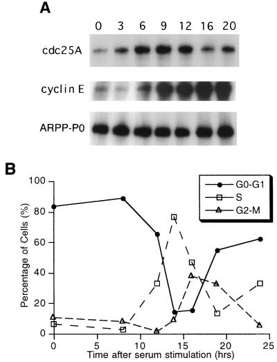
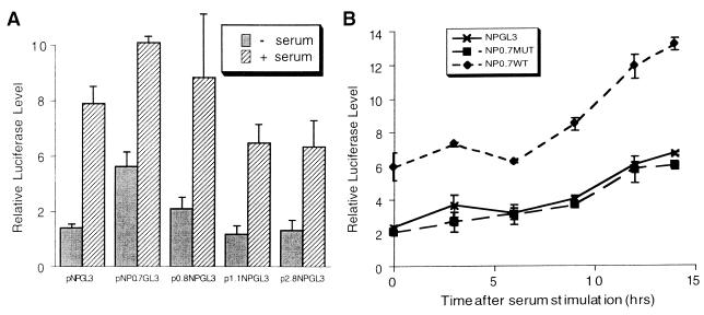
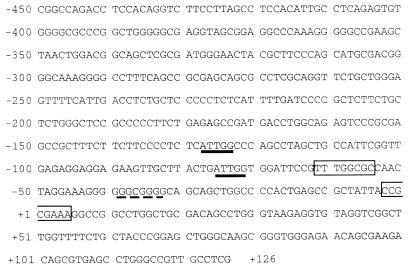
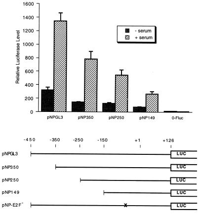
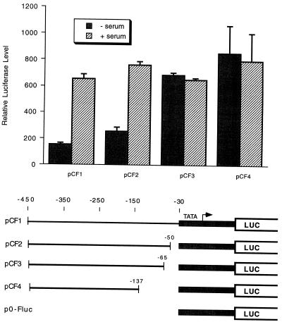
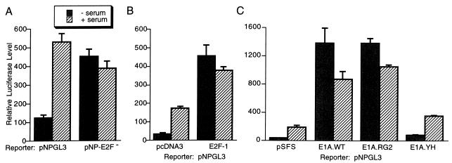
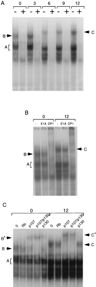
The cdc25A gene encodes a tyrosine phosphatase which activates cyclin-dependent kinase activity in the G1 phase of the cell cycle. cdc25A RNA levels are induced from 3 to 6 h after serum induction of serum-starved NIH 3T3 cells, suggesting that the cdc25A gene is a delayed-early gene. Analysis of cdc25A promoter constructs showed that the cdc25A promoter is sufficient for serum induction. Surprisingly for a gene expressed in early to mid-G1, serum induction of the promoter requires an E2F site at position -62 in the promoter. Deletion or point mutation of the E2F site resulted in activation of expression in serum-starved cells and no further induction by serum treatment. E2F factors were found to bind to the cdc25A E2F site along with the retinoblastoma protein (Rb) family members p130 and p107. A shift in mobility of the E2F-p107 complex in extracts of cells induced for 6 h correlated with induction of cdc25A expression. These results suggest that serum induction of cdc25A expression is mediated by inactivation of p107 or p130, both of which repress transcription when bound to the promoter through E2F.


Figures

FIG. 1
FIG. 1
(A) Serum induction of cdc25A mRNA in NIH 3T3 cells. NIH 3T3 cells were serum starved for 48 h and induced with serum for the time periods (in hours) indicated above the lanes. Total RNA was isolated and analyzed by RNase protection for cdc25A, cyclin E, and ARPP-P0 as described in Materials and Methods. The areas of separate gels with specifically protected bands are shown. (B) The cell cycle phase of serum-starved and -induced NIH 3T3 cells was determined by propidium iodide staining and flow cytometry.
FIG. 2
FIG. 2
Serum induction of cdc25A promoter constructs. (A) NIH 3T3 cells were transfected with the indicated cdc25A promoter-luciferase constructs containing nucleotides −450 to +126 of the cdc25A promoter, pNPGL3, and additional genomic fragments of the cdc25A gene (17). An internal control plasmid, pRL-SV40P, expressing Renilla luciferase was cotransfected. The transfected cells were serum starved (−serum) and then induced with 20% serum for 13 h (+ serum). Firefly luciferase activities were measured and normalized to the internal control Renilla luciferase activity. Shown are the averages of data from three experiments and the standard errors of the means (SEM; error bars). (B) NIH 3T3 cells were transfected with the indicated reporter genes and pRL-SV40P, serum starved, and then induced with serum for the indicated periods. The results are the average normalized luciferase activities of three experiments ± the standard errors of the means. NP0.7WT = pNP0.7GL3; NP0.7MUT contains a double point mutation in the myc binding site in NP0.7WT.
FIG. 3
FIG. 3
Sequence of the cdc25A promoter. The sequence of the human cdc25A promoter in pNPGL3 was determined. Position +1 indicates the 5′ end of the full-length cDNA (16). Two potential E2F sites are boxed, CAAT boxes are underlined, and a potential SP1 site has a dashed underline. The start codon for cdc25A is at position +460.
FIG. 4
FIG. 4
Serum induction of cdc25A promoter deletions. The constructs indicated at the bottom of the figure were transfected into NIH 3T3 cells with pRL-SV40P as an internal control and analyzed for serum induction as described in the legend to Fig. 2A. A minimal c-fos promoter plasmid, p0-Fluc (see Fig. 5), was tested as a control. Shown are the averages of data from three experiments and the standard errors of the means (error bars). − serum, no serum induction; + serum, serum induction performed.
FIG. 5
FIG. 5
Serum induction of heterologous-promoter constructs. Regions of the cdc25A promoter were fused upstream of a c-fos minimal promoter construct as indicated at the bottom of the figure (LUC, luciferase gene). The constructs were transfected into NIH 3T3 cells with pRL-SV40P and either not serum induced (− serum) or serum induced (+ serum) as described in the legend to Fig. 2A. Shown are the averages of data from three experiments and the standard errors of the means (error bars). The c-fos minimal promoter alone, p0-Fluc, was not inducible (Fig. 4).
FIG. 6
FIG. 6
E2F regulation of the cdc25A promoter. (A) The cdc25A E2F site at position −62 was mutated as diagrammed in Fig. 4 (pNP-E2F) and tested for serum induction in NIH 3T3 cells. (B) The cdc25A reporter construct pNPGL3 was transfected with an E2F-1 expression vector or vector pcDNA3 and tested for serum-induced expression. (C) Adenovirus E1A expression vectors were transfected with pNPGL3 and pRL-SV40P. Wild-type E1A (E1A.WT) and mutants were transfected. A plasmid with a frameshift of E1A, pSFS, which does not express E1A served as a control vector. The E1A.RG2 mutant binds Rb family proteins but not p300 or CBP (57). The E1A YH47/928 mutant (E1A.YH) binds p300 and CBP but not Rb family proteins (57). Shown are the averages of data from three experiments and the standard errors of the means (error bars). − serum, no serum induction; + serum, serum induction performed.
FIG. 7
FIG. 7
Binding of E2F and Rb family proteins to the cdc25A E2F site. (A) Extracts were prepared from NIH 3T3 cells that were serum starved and then induced with serum for the time periods (in hours) indicated above the lanes. Extracts were incubated with 32P-labelled cdc25A E2F site oligonucleotide with a 40-fold excess of unlabelled probe (+) or nonspecific competitor (−). (B) Extracts from serum-starved (0) or 12-h serum-stimulated (12) cells were incubated with the E2F site probe, as described for panel A, without antiserum (−), with a nonspecific antiserum (anti-E1A), or with anti-DP1. (C) Extracts from serum-starved (0) or 12-h serum-stimulated (12) cells were incubated with antiserum to Rb, p107, or p130 or with a combination of anti-p107 and anti-p130 sera as indicated. Complexes A, B, and C indicate E2F containing the complexes discussed in the text. B’ and C’ indicate complexes supershifted by antiserum.

References

    1. Beckmann H, Su L K, Kadesch T. TFE3: a helix-loop-helix protein that activates transcription through the immunoglobulin enhancer muE3 motif. Genes Dev. 1990;4:167–179. - PubMed
    1. Beijersbergen R L, Carlee L, Kerkhoven R M, Bernards R. Regulation of the retinoblastoma protein-related p107 by G1 cyclin complexes. Genes Dev. 1995;9:1340–1353. - PubMed
    1. Beijersbergen R L, Kerkhoven R M, Zhu L, Carlee L, Voorhoeve P M, Bernards R. E2F-4, a new member of the E2F gene family, has oncogenic activity and associates with p107 in vivo. Genes Dev. 1994;8:2680–2690. - PubMed
    1. Botz J, Zerfass-Thome K, Spitkovsky D, Delius H, Vogt B, Eilers M, Hatzigeorgiou A, Jansen-Dürr P. Cell cycle regulation of the murine cyclin E gene depends on an E2F binding site in the promoter. Mol Cell Biol. 1996;16:3401–3409. - PMC - PubMed
    1. Bush A, Mateyak M, Dugan K, Obaya A, Adachi S, Sedivy J, Cole M. c-myc null cells misregulate cad and gadd45 but not other proposed c-Myc targets. Genes Dev. 1998;12:3797–3802. - PMC - PubMed

Publication types

MeSH terms

Substances

LinkOut - more resources