. 1999 Jul;19(7):4729-38.
doi: 10.1128/MCB.19.7.4729.

p70(s6k) integrates phosphatidylinositol 3-kinase and rapamycin-regulated signals for E2F regulation in T lymphocytes

Affiliations

p70(s6k) integrates phosphatidylinositol 3-kinase and rapamycin-regulated signals for E2F regulation in T lymphocytes

P Brennan et al. Mol Cell Biol. 1999 Jul.

Abstract

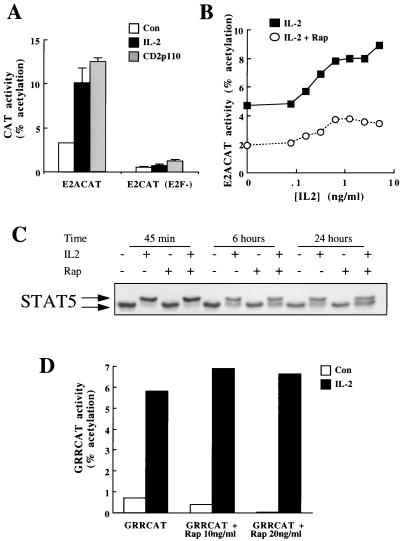
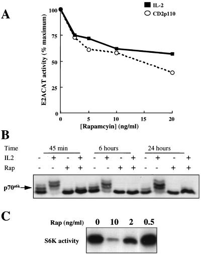
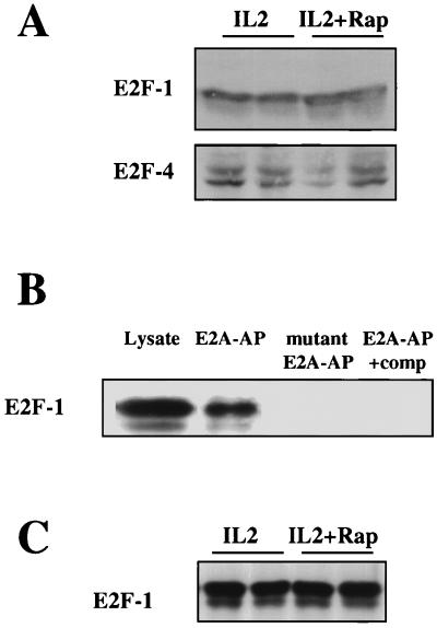
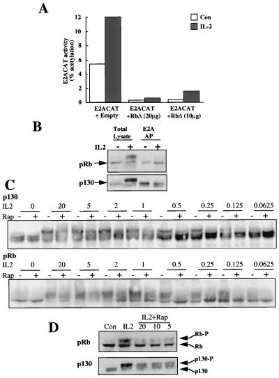
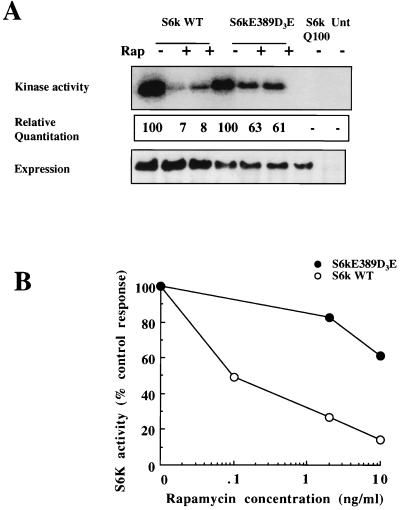
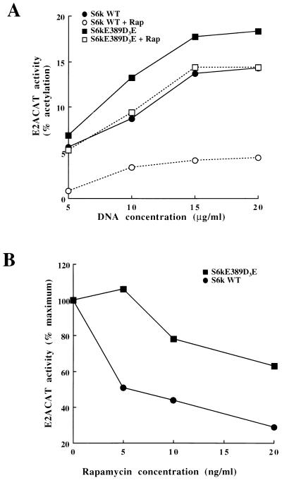
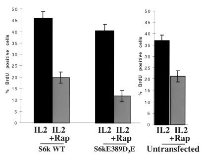
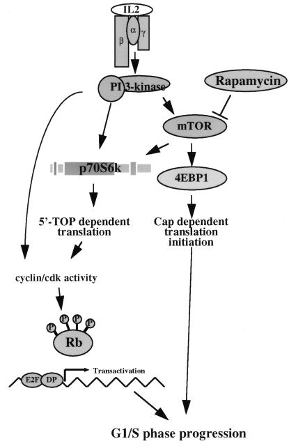
In T lymphocytes, the hematopoietic cytokine interleukin-2 (IL-2) uses phosphatidylinositol 3-kinase (PI 3-kinase)-induced signaling pathways to regulate E2F transcriptional activity, a critical cell cycle checkpoint. PI 3-kinase also regulates the activity of p70(s6k), the 40S ribosomal protein S6 kinase, a response that is abrogated by the macrolide rapamycin. This immunosuppressive drug is known to prevent T-cell proliferation, but the precise point at which rapamycin regulates T-cell cycle progression has yet to be elucidated. Moreover, the effects of rapamycin on, and the role of p70(s6k) in, IL-2 and PI 3-kinase activation of E2Fs have not been characterized. Our present results show that IL-2- and PI 3-kinase-induced pathways for the regulation of E2F transcriptional activity include both rapamycin-resistant and rapamycin-sensitive components. Expression of a rapamycin-resistant mutant of p70(s6k) in T cells could restore rapamycin-suppressed E2F responses. Thus, the rapamycin-controlled processes involved in E2F regulation appear to be mediated by p70(s6k). However, the rapamycin-resistant p70(s6k) could not rescue rapamycin inhibition of T-cell cycle entry, consistent with the involvement of additional, rapamycin-sensitive pathways in the control of T-cell cycle progression. The present results thus show that p70(s6k) is able to regulate E2F transcriptional activity and provide direct evidence for the first time for a link between IL-2 receptors, PI 3-kinase, and p70(s6k) that regulates a crucial G1 checkpoint in T lymphocytes.


Figures

FIG. 1
FIG. 1
Rapamycin inhibits IL-2 activation of E2ACAT. (A) Quiescent Kit225 cells (1.5 × 10⁷ per sample) were cotransfected with E2ACAT or the mutated reporter [E2CAT(E2F)] and either empty vector or CD2p110 (active PI 3-kinase) (20 μg). After 2 h, cells transfected with empty vector were stimulated with IL-2 (20 ng/ml). Open bars, control (Con); solid bars, IL-2; shaded bars, CD2p110. After 22 h, samples were harvested and lysed, and CAT activity was measured. (B) Quiescent Kit225 cells (1.5 × 10⁷ per sample) were transfected with E2ACAT (20 μg). Cells were treated with 20 ng of rapamycin/ml (open circles) or left untreated (solid squares) for 20 min prior to stimulation with various doses of IL-2 as indicated. After 18 h, samples were harvested and assayed for CAT activity. (C) Kit225 cells (2 × 10⁶ per ml; 5 ml per sample) were pretreated with rapamycin (20 ng/ml) and incubated with IL-2 (20 ng/ml) as indicated for 45 min, 6 h, and 24 h. Total cell lysates were generated and resolved by SDS-PAGE, and Western blotting was performed. Protein was detected by using antibodies specific for STAT5. (D) Quiescent Kit225 cells (1.5 × 10⁷ per sample) were transfected with GRRCAT (20 μg). Cells were pretreated with rapamycin (20 ng/ml) for 20 min prior to stimulation with IL-2 (20 ng/ml) as indicated. After 18 h, samples were harvested and assayed for CAT activity.
FIG. 2
FIG. 2
Rapamycin effects on E2ACAT and p70s6k. (A) Quiescent Kit225 cells (1.5 × 10⁷ per sample) were transfected with E2ACAT (20 μg) and with either 20 μg of empty vector (solid squares) or pEF rCD2p110 (active PI 3-kinase) (open circles). Cells were left for 4 h. They were pretreated with rapamycin (20 ng/ml), and the cells transfected with empty vector were stimulated with IL-2 (20 ng/ml). After 18 h, samples were harvested and assayed for CAT activity. Data are expressed as percentages of maximum activity, with 100% representing 15% ± 3% acetylation for IL-2 and 24% ± 4% acetylation for cells cotransfected with CD2p110. (B) Kit225 cells (106 per ml; 5 ml per sample) were pretreated with rapamycin (20 ng/ml) and incubated with IL-2 (20 ng/ml) as indicated for 45 min, 6 h, and 24 h. Total cell lysates were generated and resolved by SDS-PAGE, and Western blotting was performed. Protein was detected by using antibodies specific for S6 kinase. (C) Kit225 cells (1.5 × 10⁷ per sample) were transfected with an expression vector for myc epitope-tagged p70s6k. Cells were left overnight and treated for 20 min with rapamycin at the doses indicated. Cells were lysed, the expressed p70s6k was immunoprecipitated with myc tag-specific antibody, and kinase activity was measured. S6 substrate phosphorylation for S6 kinase assays was analyzed by autoradiography.
FIG. 3
FIG. 3
Rapamycin and E2F. (A) Kit225 cells (106 per ml; 5 ml per sample) were pretreated with rapamycin (Rap) (20 ng/ml) and incubated with IL-2 (20 ng/ml) as indicated for 20 h. Total cell lysates were generated and resolved by SDS-PAGE, and Western blotting was performed. Protein was detected by using specific antibodies for E2F-1 and E2F-4. (B) Kit225 cells (106 per ml; 10 ml per sample) were lysed, and DNA affinity precipitations were performed with biotinylated oligonucleotides containing E2A sequence (E2A-AP), with a mutant E2A biotinylated oligonucleotide (two point mutations in each E2F binding site) (mutant E2A-AP) or with biotinylated oligonucleotides containing E2A sequence in the presence of a 10-fold excess of unbiotinylated E2F binding oligonucleotide as a competitor (E2A-AP+comp). Samples were analyzed by SDS-PAGE and Western blotting. Protein was detected with E2F-1-specific antibodies. (C) Kit225 cells (106 per ml; 10 ml per sample) were pretreated with rapamycin (20 ng/ml) and stimulated with IL-2 (20 ng/ml) for 20 h. Samples were lysed, and DNA affinity precipitations were performed with biotinylated oligonucleotides containing E2A sequence. Samples were analyzed by SDS-PAGE and Western blotting. Protein was detected with E2F-1-specific antibodies.
FIG. 4
FIG. 4
Rapamycin and pocket proteins. (A) Quiescent Kit225 cells (1.5 × 10⁷ per sample) were cotransfected with E2ACAT reporter with a mammalian expression vector for a form of pRb in which the cdc2 phosphorylation sites had been mutated (pRbΔ) or with empty vector as a control. After 4 h, the cells were left untreated (Con) or stimulated with IL-2 (20 ng/ml). Eighteen hours later, the cells were harvested, lysed, and assayed for CAT. (B) Peripheral blood lymphocytes (106 per ml; 10 ml per sample) were incubated with IL-2 (20 ng/ml) as indicated for 20 h. Samples were lysed, and DNA affinity precipitations (AP) were performed with biotinylated oligonucleotides containing E2A sequence. Samples were analyzed by SDS-PAGE and Western blotting. Protein was detected with specific pRb and p130 antibodies. (C) Peripheral blood lymphocytes (106 per ml; 5 ml per sample) were pretreated with rapamycin (20 ng/ml) and stimulated with IL-2 at the doses indicated (in nanograms per milliliter) for 20 h. Total cell lysates were generated and resolved by SDS-PAGE, and Western blotting was performed. Protein was detected by using specific antibodies for pRb and p130. (D) Peripheral blood lymphocytes (106 per ml; 5 ml per sample) were pretreated with various doses of rapamycin as indicated (in nanograms per milliliter) and stimulated with IL-2 (20 ng/ml) for 20 h. Total cell lysates were generated and resolved by SDS-PAGE, and Western blotting was performed. Protein was detected with specific antibodies for pRb and p130. Con, control.
FIG. 5
FIG. 5
p70s6k increases E2F transcriptional activity. (A) (Top) Quiescent Kit225 cells (1.5 × 10⁷ per sample) were transfected with E2ACAT (20 μg) and with 10 μg each of either empty vector, wild-type p70s6k (S6k WT), or p70s6kQ100 (Q100) (kinase-dead S6k). Cells were left for 4 h and were either stimulated with 20 ng of IL-2/ml (solid bars) or left unstimulated (open bars) (Con). After 18 h, samples were harvested and assayed for CAT activity. (Bottom) In parallel experiments, Kit225 cells (1.5 × 10⁷ per sample) were transfected with an expression vector for myc epitope-tagged S6k constructs. Cells were left overnight and were lysed, and the expressed S6k was immunoprecipitated with myc tag-specific antibody and separated by SDS-PAGE. Transfected myc-tagged S6k was detected with myc tag-specific antibody (9E10). (B) Quiescent Kit225 cells (1.5 × 10⁷ per sample) were transfected with E2ACAT (20 μg) and with 20 μg of wild-type p70s6k or empty vector. Cells were left for 4 h. They were then treated with various doses of rapamycin. After 18 h, samples were harvested and assayed for CAT activity.
FIG. 6
FIG. 6
p70s6kD3E-E389 is rapamycin resistant in T-cells. (A) Kit225 cells (1.5 × 10⁷ per sample) were transfected with 30 μg each of empty vector, wild-type p70s6k (S6k WT), the rapamycin-resistant mutant p70s6kD3E-E389, or the kinase-dead mutant p70s6kQ100. Cells were left overnight and treated for 20 min with rapamycin (20 ng/ml). Cells were lysed, the expressed S6k was immunoprecipitated with myc tag-specific antibody, and kinase activity was measured. S6 substrate phosphorylation for S6 kinase assays was analyzed by autoradiography (top), and expression was measured by Western blot analysis (bottom). (Center) Incorporated radioactivity was measured with a PhosphorImager. Data are expressed relative to activity in the absence of rapamycin, taken as 100. (B) Kit225 cells (1.5 × 10⁷ per sample) were transfected with 30 μg each of empty vector, wild-type p70s6k, p70s6kD3E-E389, or p70s6kQ100. Cells were left overnight and treated for 20 min with rapamycin at the doses indicated. Cells were lysed, the expressed S6k was immunoprecipitated with myc tag-specific antibody, and kinase activity was measured. S6 substrate-incorporated radioactivity was measured with a PhosphorImager. Data are expressed relative to activity in the absence of rapamycin, taken as 100%.
FIG. 7
FIG. 7
p70s6kD3E-E389 rescues rapamycin inhibition of E2F transcriptional activity. (A) Quiescent Kit225 cells (1.5 × 10⁷ per sample) were transfected with E2ACAT (20 μg) and with wild-type (WT) p70s6k or the rapamycin-resistant mutant (p70s6kD3E-E389) at various concentrations. Cells were left for 4 h and then treated with rapamycin (Rap) (20 ng/ml). After 18 h, samples were harvested and assayed for CAT activity. (B) Quiescent Kit225 cells (1.5 × 10⁷ per sample) were transfected with E2ACAT (20 μg) and with wild-type (WT) p70s6k or the rapamycin-resistant mutant (p70s6kD3E-E389). Cells were left for 4 h. They were then pretreated for 20 min with various doses of rapamycin and were stimulated with IL-2 (20 ng/ml). After 18 h, samples were harvested and assayed for CAT activity.
FIG. 8
FIG. 8
p70s6kD3E-E389 does not rescue rapamycin inhibition of T-cell cycle entry. Quiescent Kit225 cells (1.5 × 10⁷ per sample) were transfected with wild-type (WT) p70s6k or p70s6kD3E-E389. Cells were left overnight. They were then pretreated for 20 min with rapamycin (Rap) (20 ng/ml) and stimulated with IL-2 (20 ng/ml). After 8 h, 10 μM BrdU was added to samples. Twelve hours later, cells were harvested and assayed for BrdU incorporation and protein expression by FACS.
FIG. 9
FIG. 9
IL-2 signaling to E2F and the cell cycle. Role of p70s6k in E2F transactivation in T cells. Our model places p70s6k upstream of translation events leading to E2F transcriptional activity but demonstrates that other rapamycin-sensitive signals are required for cell cycle progression.

References

    1. Beadling C, Guschin D, Witthuhn B A, Ziemiecki A, Ihle J N, Kerr I M, Cantrell D A. Activation of JAK kinases and STAT proteins by interleukin-2 and interferon-α, but not the T cell antigen receptor, in human T lymphocytes. EMBO J. 1994;13:5605–5615. - PMC - PubMed
    1. Beadling C, Ng J, Babbage J W, Cantrell D A. Interleukin-2 activation of STAT5 requires the convergent action of tyrosine kinases and a serine/threonine kinase distinct from the Raf-1/Erk2 MAP kinase pathway. EMBO J. 1996;15:1902–1913. - PMC - PubMed
    1. Beretta L, Gingras A C, Svitkin Y V, Hall M N, Sonenberg N. Rapamycin blocks the phosphorylation of 4E-BP1 and inhibits cap-dependent initiation of translation. EMBO J. 1996;15:658–664. - PMC - PubMed
    1. Botz J, Zerfass-Thome K, Spitzovsky D, Delius H, Vogt B, Eilers M, Hatzigeorgiou A, Jansen-Durr P. Cell cycle regulation of the murine cyclin E gene depends on an E2F binding site in the promoter. Mol Cell Biol. 1996;16:3401–3409. - PMC - PubMed
    1. Brennan P, Babbage J W, Burgering B M T, Groner B, Reif K, Cantrell D A. Phosphatidylinositol 3-kinase controls E2F transcriptional activity in response to interleukin-2. Immunity. 1997;7:679–689. - PubMed

Publication types

MeSH terms